Development
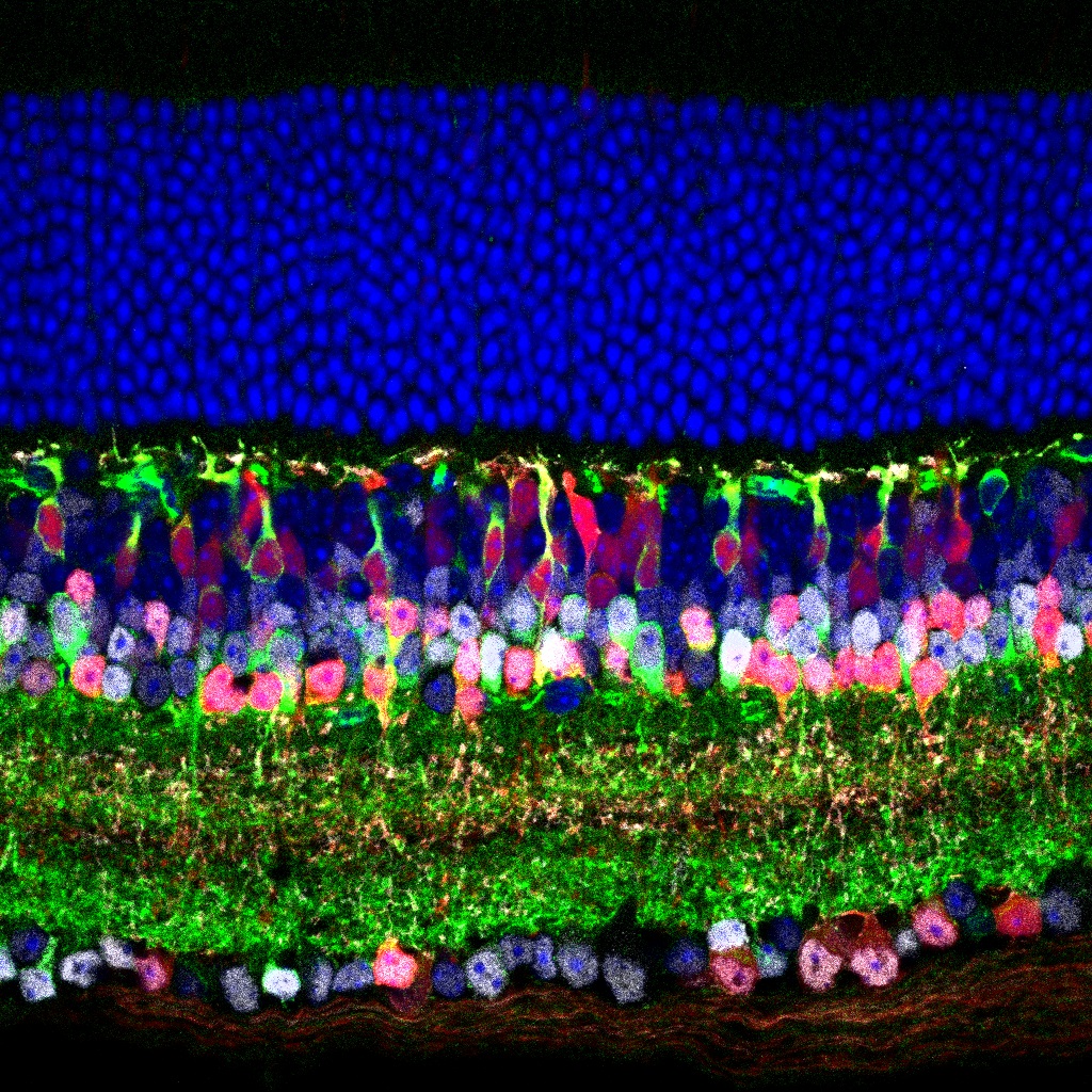
This image shows the cellular layers of an adult mouse retina, stained for markers of amacrine cells and type 3b bipolar cells.
In this study, authors find an important new locus of compensatory synaptic strength changes in the early developing chick autonomic nervous system.
This paper investigates spatial and temporal patterns of key enzymes involved in purine synthesis.

Dr. Matthew Colonnese gives behind-the-scenes details about his eNeuro paper and cogitates on the implications of his surprising findings, in an episode of the webinar series SfN Journals: In Conversation. Here we provide a teaser for the episode available to watch on-demand.
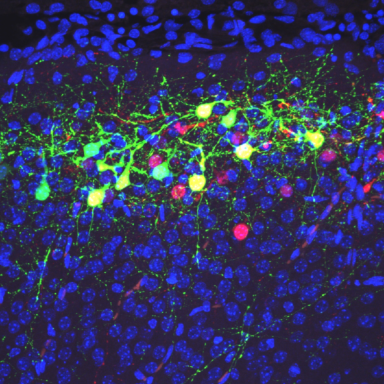
Green cells in this image are neurons distributed in the superficial region of the cerebral neocortex at postnatal day 9 in a heterozygous mutant mouse of Dab1.
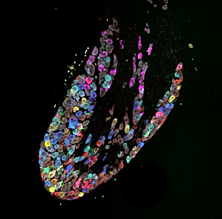
A composite of 5 images captured following each of 5 rounds of labeling and reprobing for a total of 12 mRNA transcripts and neuronal marker.
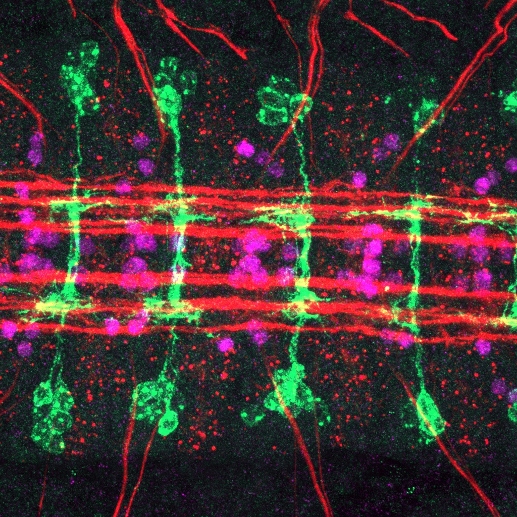
This image shows the developing central nervous system in a Drosophila embryo.
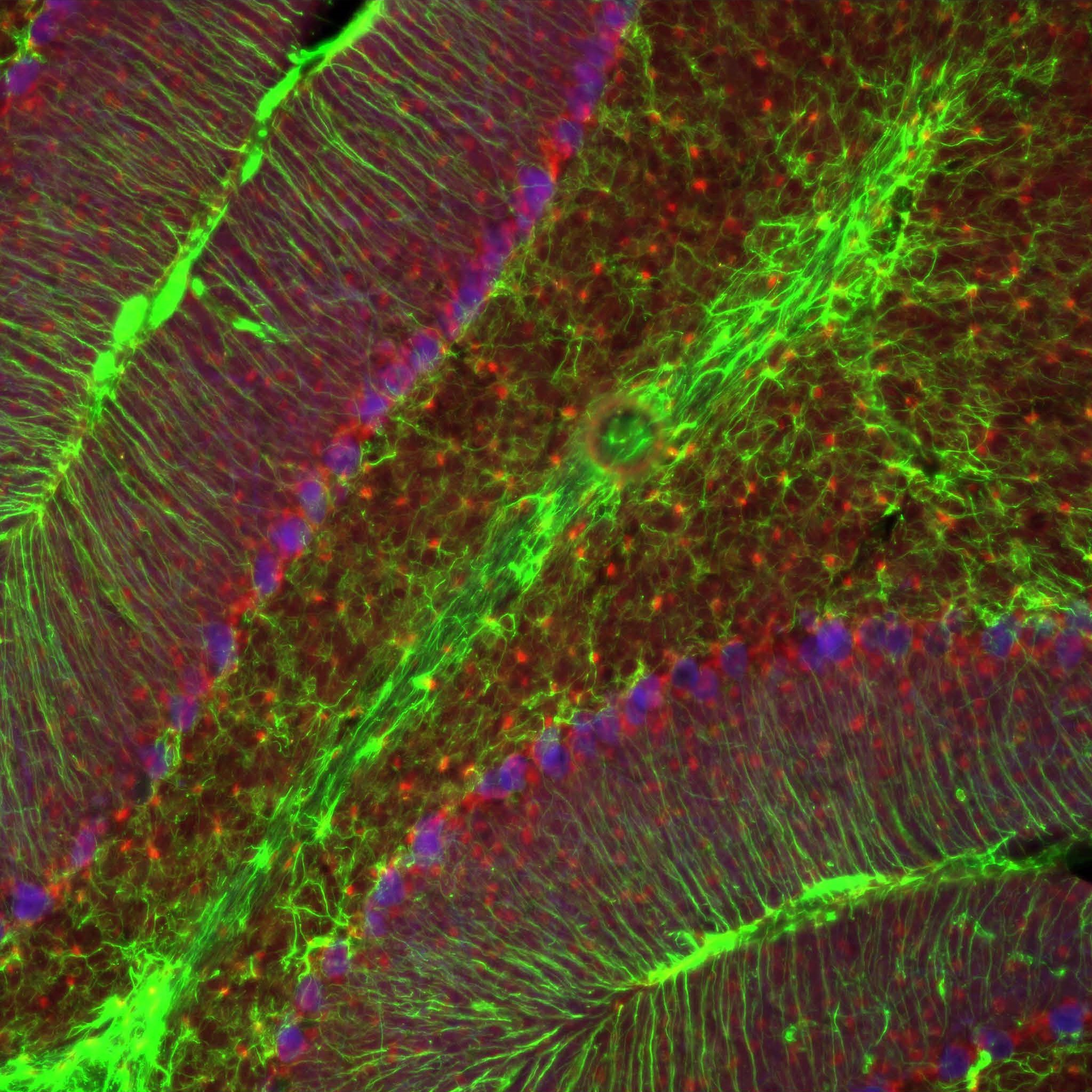
Part of the juvenile cerebellar cortex of a mouse at postnatal day 21.
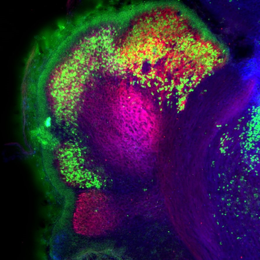
Purkinje cell clusters in the mouse cerebellum at embryonic day 17.5.
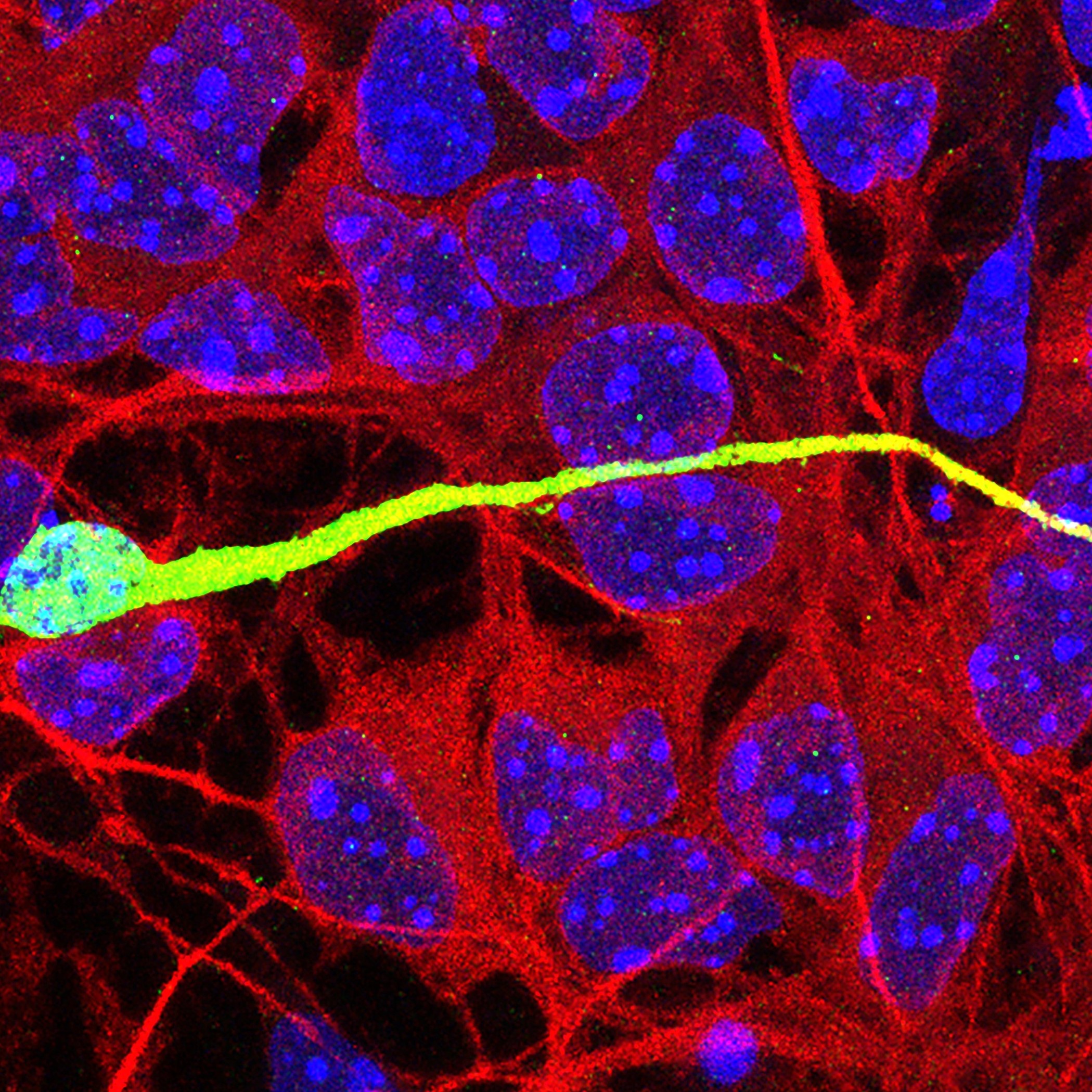
This image shows an interneuron migrating on a monolayer of cortical feeder cells in an in vitro co-culture assay.
FOLLOW US
 RSS Feed
TAGS
CATEGORIES


