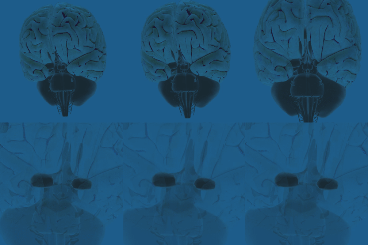
In the 1970s, anthropologist Paul Ekman proposed that humans experienced six basic emotions: anger, fear, surprise, disgust, joy, and sadness. Since then, scientists have disputed the exact number of human emotions — some researchers maintain there are only four, while others count as many as 27. And, scientists also debate whether they are universal to all human cultures and whether we’re born with them or learn them through experience. Even the definition of emotion is a topic of controversy. One thing is clear though — emotions arise from activity in distinct regions of the brain.
Three brain structures appear most closely linked with emotions: the amygdala, the insula or insular cortex, and a structure in the midbrain called the periaqueductal gray.
A paired, almond-shaped structure deep within the brain, the amygdala integrates emotions, emotional behavior, and motivation. It interprets fear, helps distinguish friends from foes, and identifies social rewards and how to attain them. The amygdala is also important for a type of learning called classical conditioning. Russian physiologist Ivan Pavlov first described classical conditioning, where, through repeated exposure, a stimulus elicits a particular response, in his studies of digestion in dogs. The dogs salivated when a lab technician brought them food. Over time, Pavlov noted the dogs also began to salivate at the mere sight of the technician, even if he was empty-handed.
The insula is the source of disgust — a strong negative reaction to an unpleasant odor, for instance. The experience of disgust may protect you from ingesting poison or spoiled food. Studies using magnetic resonance imaging (MRI) have found the insula lights up with activity when someone feels or anticipates pain. Neuroscientists think the insula receives a status report about the body’s physiological state and generates subjective feelings about it thus linking internal states, feelings, and conscious actions.
The periaqueductal gray, located in the brainstem, has also been implicated in pain perception. It contains receptors for pain-reducing compounds like morphine and oxycodone, and can help quell activity in pain-sensing nerves — it might be part of the reason you can sometimes distract yourself from pain so you don’t feel it as acutely. The periaqueductal gray is also involved in defensive and reproductive behaviors, maternal attachment, and anxiety.
While emotions are intangible and hard to describe — even for scientists — they serve important purposes, helping us learn, initiate actions, and survive.
Adapted from the 8th edition of Brain Facts by Deborah Halber.
CONTENTPROVIDEDBY
BrainFacts/SfN
References
Linnman C, Moulton EA, Barmettler G, Becerra L, Borsook D. 2012. Neuroimaging of the Periaqueductal Gray: State of the Field. Neuroimage. 60(1):505–522. doi:10.1016/j.neuroimage.2011.11.095.


