Snapshots in Neuroscience: Blood Vessels
This image has been selected to showcase the art that neuroscience research can create.
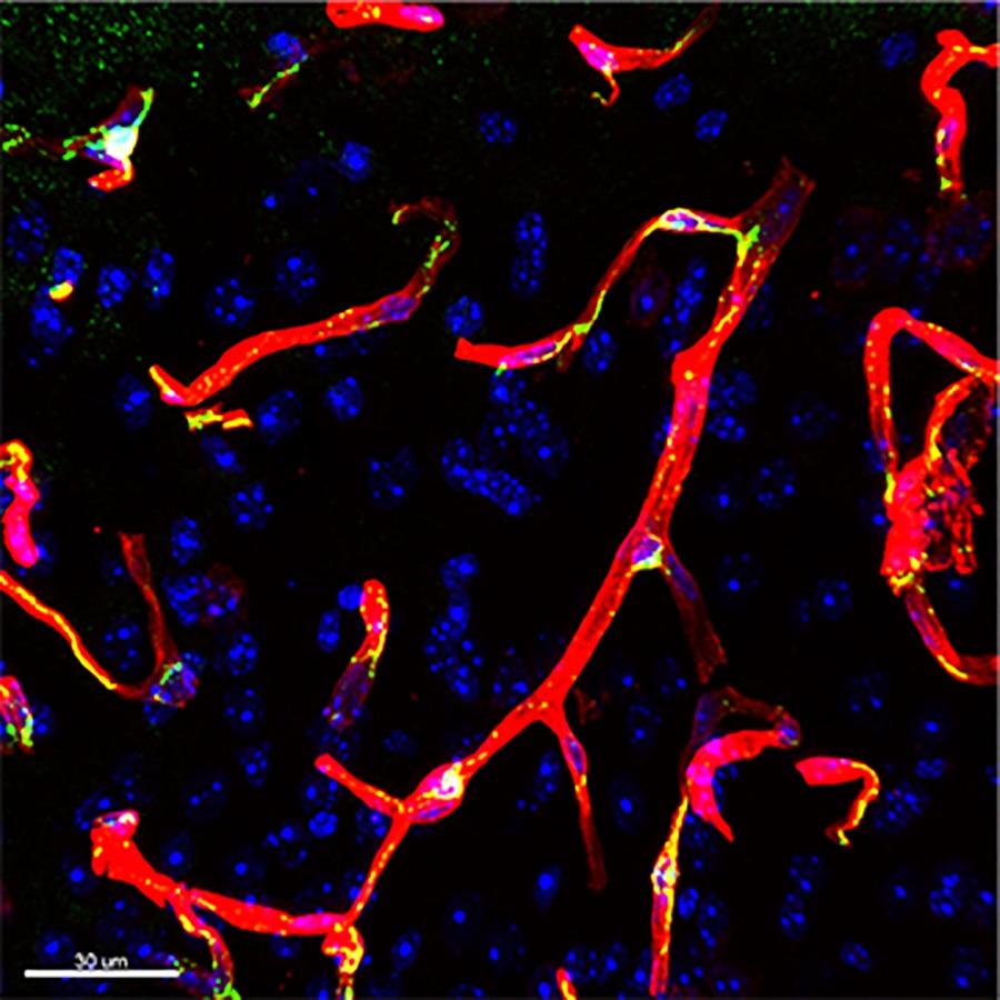
As described by Dr. Gareth Howell and colleagues: the image shows blood vessels visualized with an antibody against laminin (red) in a region of the parietal cortex from a 12-month-old mouse fed a regular diet. Also shown are pericytes (using an antibody against PDGFRβ, green) and cell nuclei (DAPI, blue).
In this study, Dr. Leah Graham showed that global deletion of an initiating factor of the complement cascade, part of the innate immune response, prevented the loss of laminin and pericytes in mice fed a western diet from 2 to 12 months old. Furthermore, obesity-induced microglia phagocytosis and breakdown of myelin in the corpus callosum were also prevented by deficiency of C1QA. Collectively, these data show that C1QA is necessary for damage to the cerebrovasculature and white matter damage in diet-induced obesity.
Read the full article:
Deficiency of Complement Component C1Q Prevents Cerebrovascular Damage and White Matter Loss in a Mouse Model of Chronic Obesity
Leah C. Graham, Heidi E. Kocalis, Ileana Soto, and Gareth R. Howell
FOLLOW US
 RSS Feed
POPULAR POSTS
TAGS
CATEGORIES


