Snapshots in Neuroscience
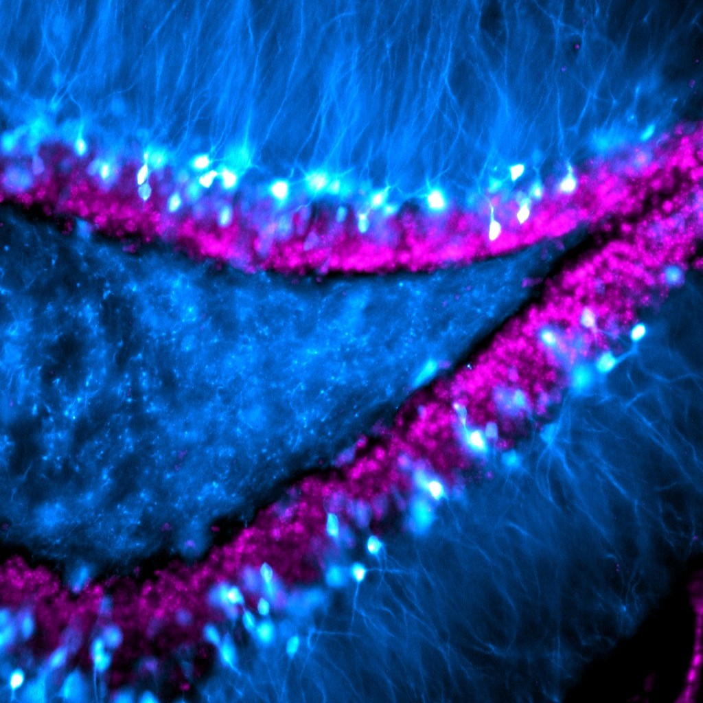
Granule cells in the dentate gyrus of a mouse, with visible extensions of their dendrites reaching out into the molecular layer.
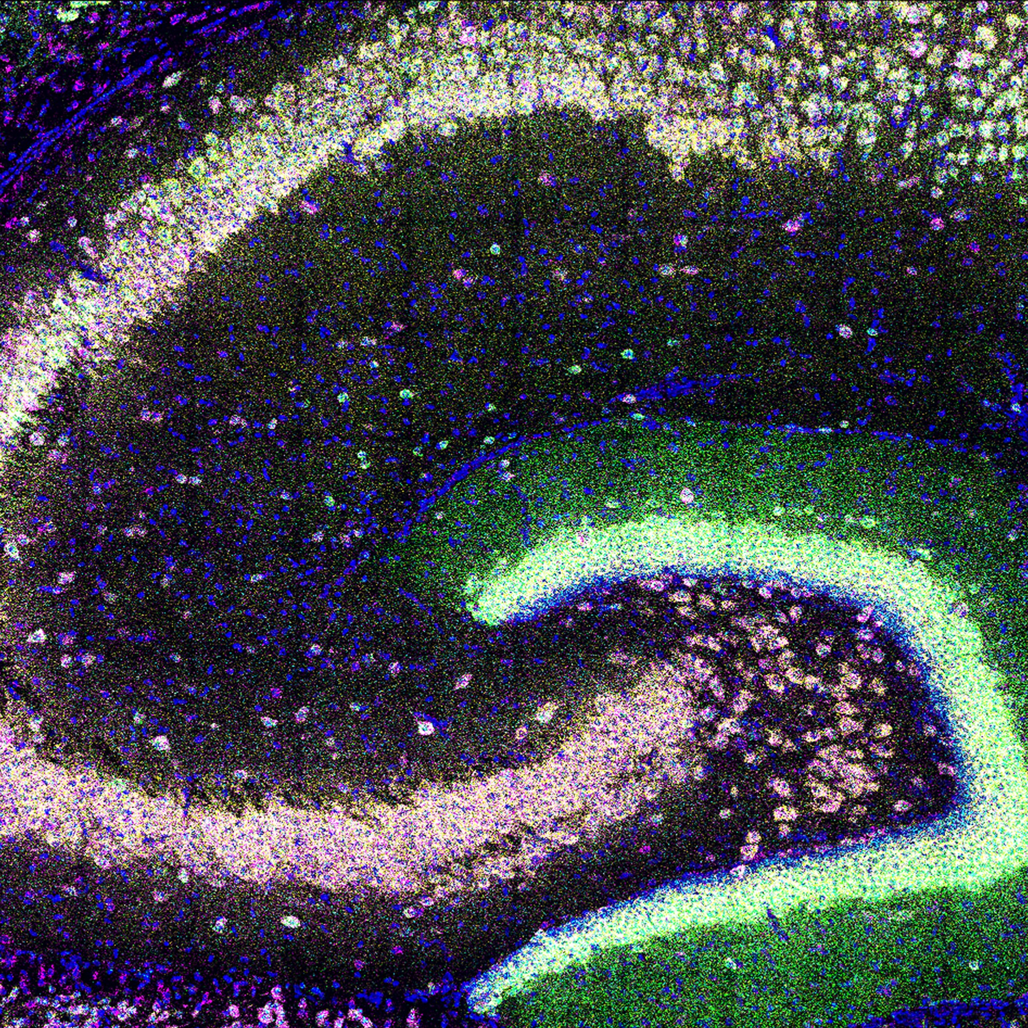
Expression of four neuropil-localized mRNA in hippocampal subregions of a young mouse.
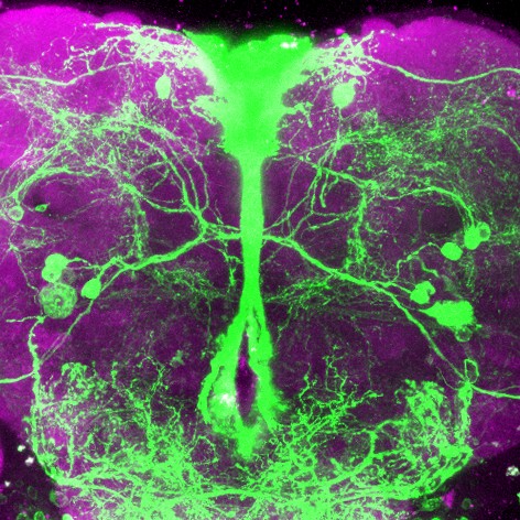
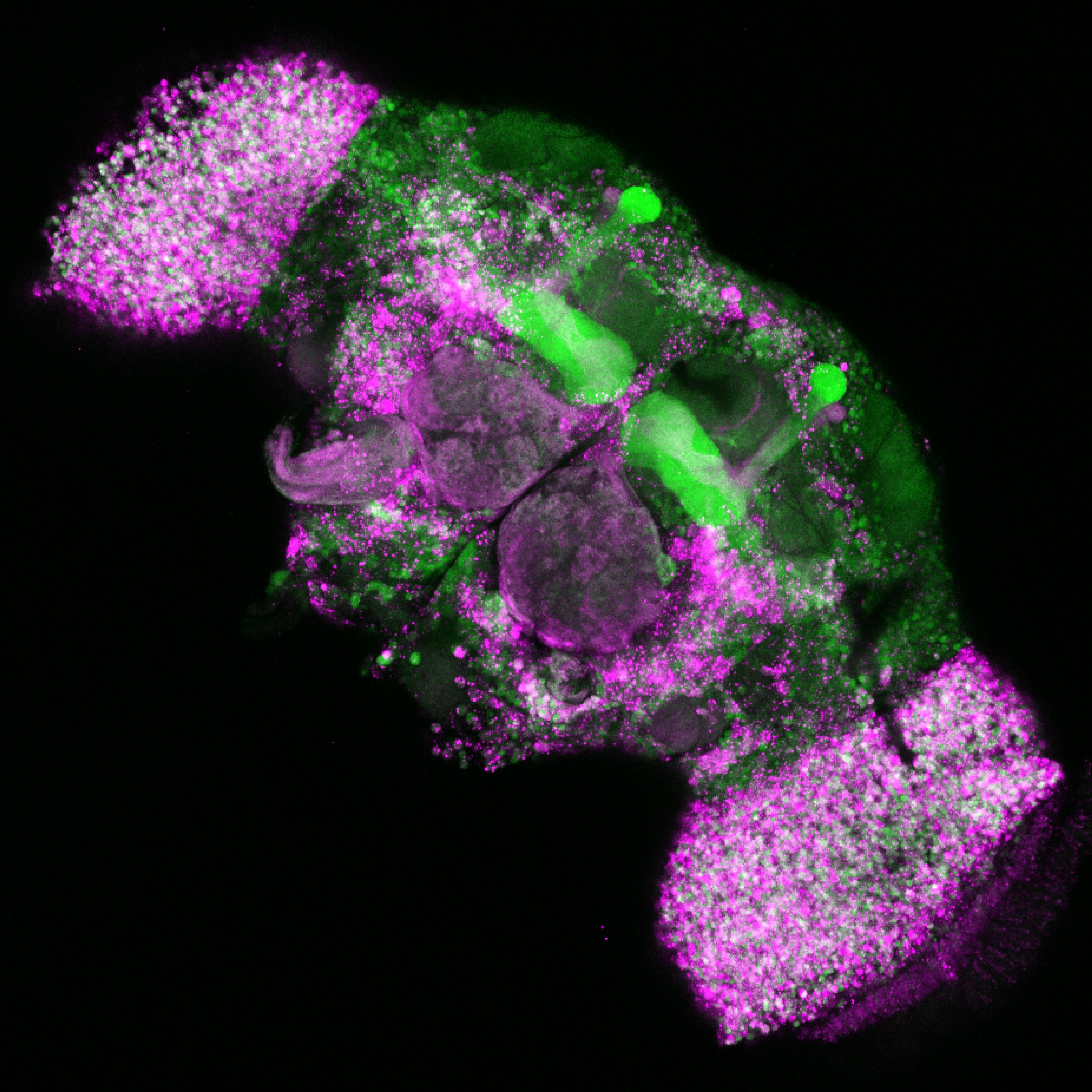
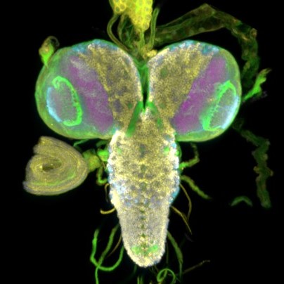
Mapping neuropeptide F receptor expressing-neurons and their role in thirsty water seeking in the adult male fruit fly.
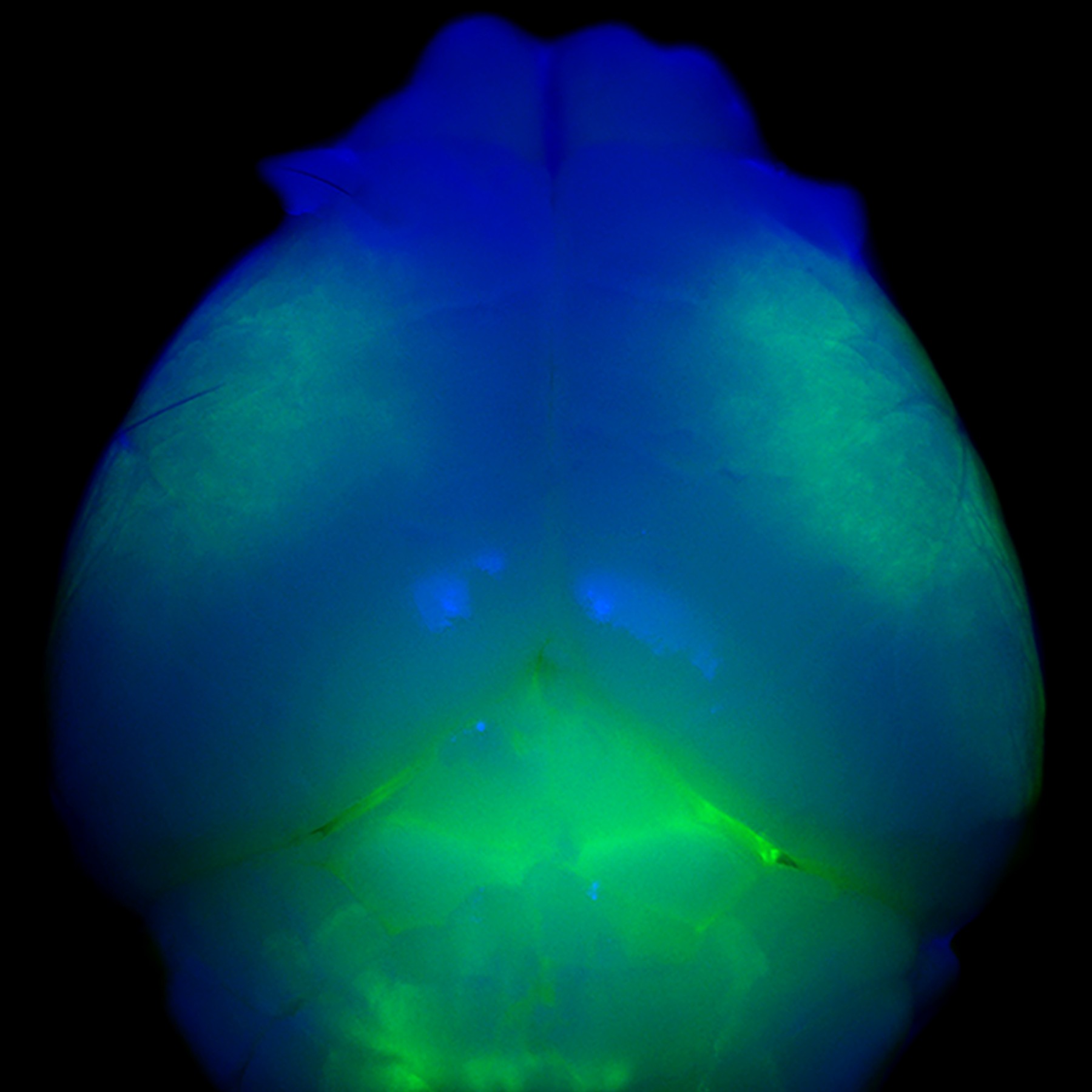
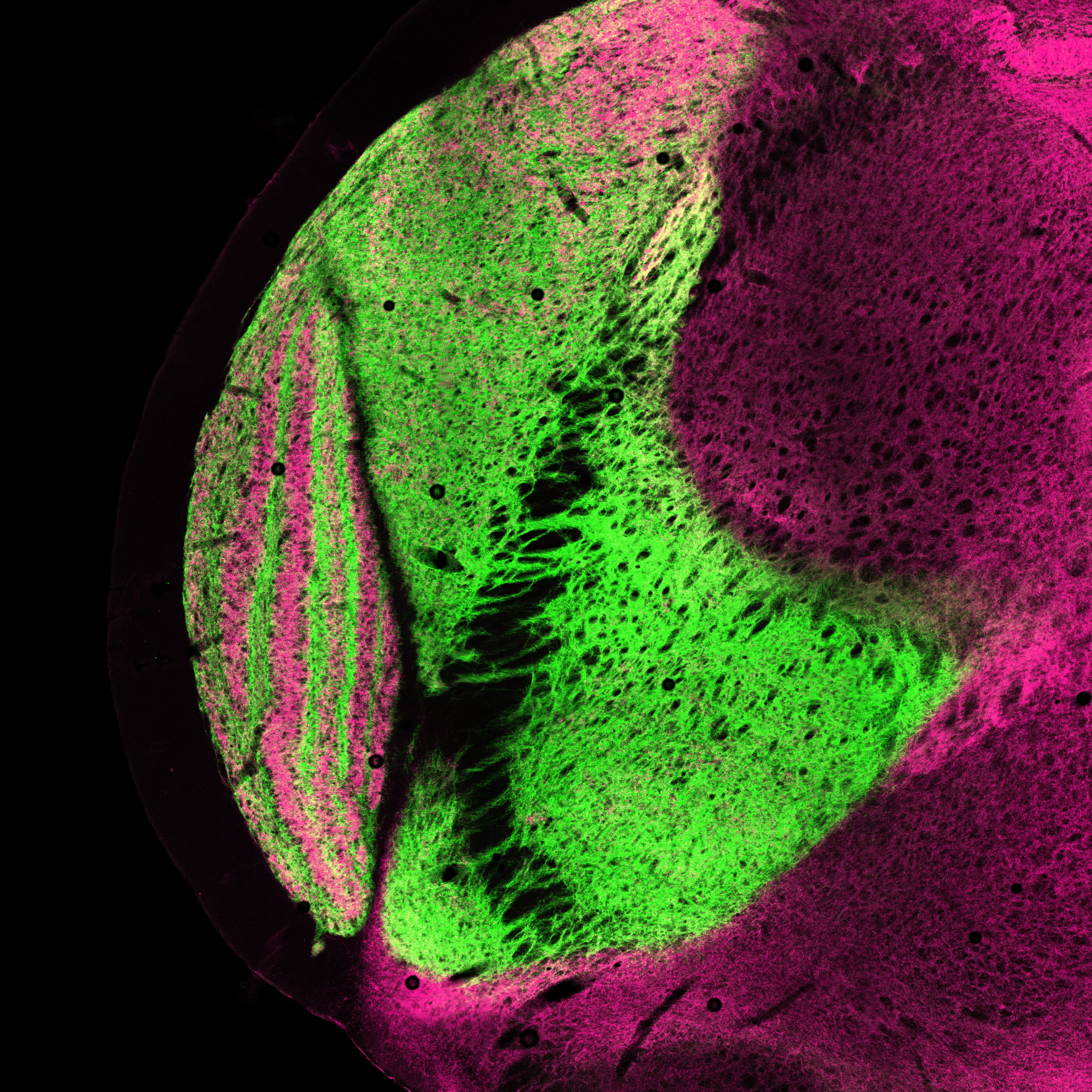
Whole mount view of a young mouse brain with all cortical layer 5 neurons expressing dystonia-related gene Klhl14, highlighted in green.
Neurons in the adult fruit fly brain have different biases in the splicing of a calcium channel that enables neurotransmission.
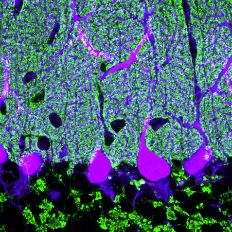
Mouse cerebellar section with a single layer of Purkinje cells extending their elaborate dendritic branches upwards into the dense molecular layer.
Excitatory axon terminals expressing vesicular glutamate transporters in the thalamus of a tree shrew.
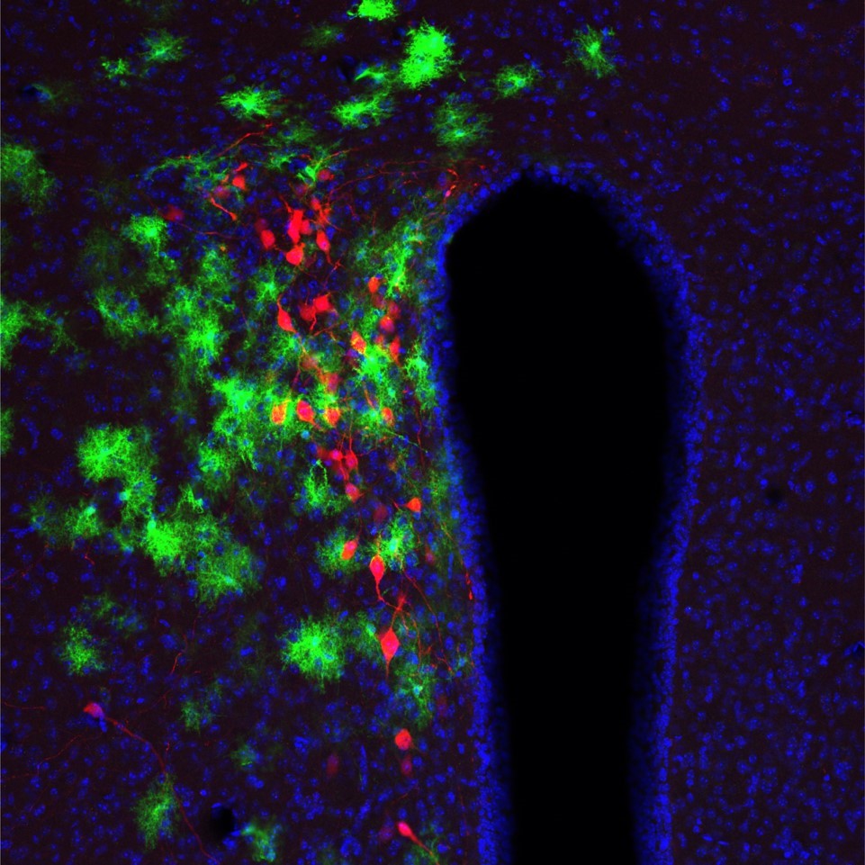
Real-time calcium dynamics of astrocytes and oxytocin neurons in the mouse paraventricular nucleus of the hypothalamus.
Exploring the neuronal expression of seizure-associated proteins in the fruit fly brain.
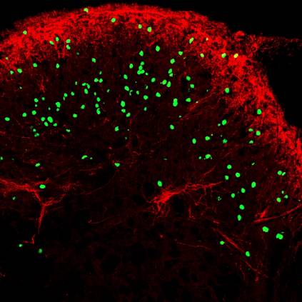
New mouse model for inducing targeted gene expression within neurons of the spinal cord dorsal horn.
FOLLOW US
TAGS
CATEGORIES


 RSS Feed


