Neuroscience Research
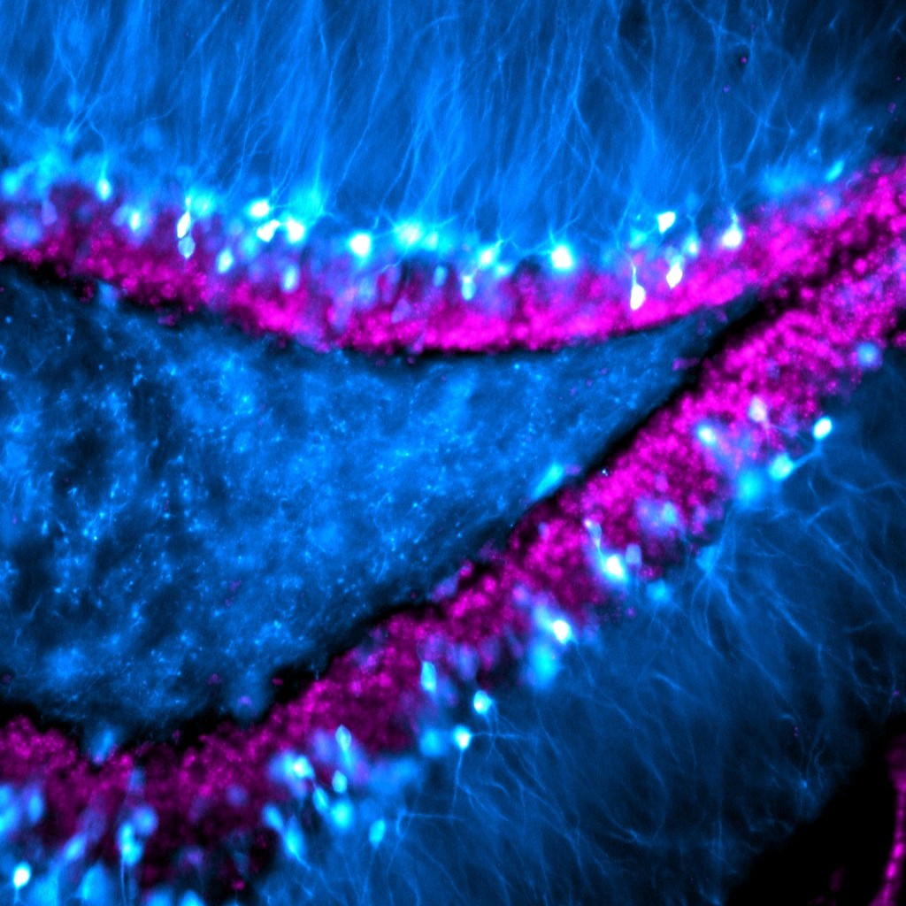
Granule cells in the dentate gyrus of a mouse, with visible extensions of their dendrites reaching out into the molecular layer.
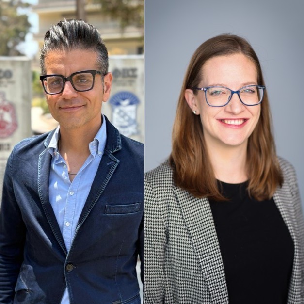
Giordano de Guglielmo and Michelle Doyle detail the challenges of creating a preclinical Alcohol Biobank. They also emphasize how refreshing and motivating it can be to work with new researchers.
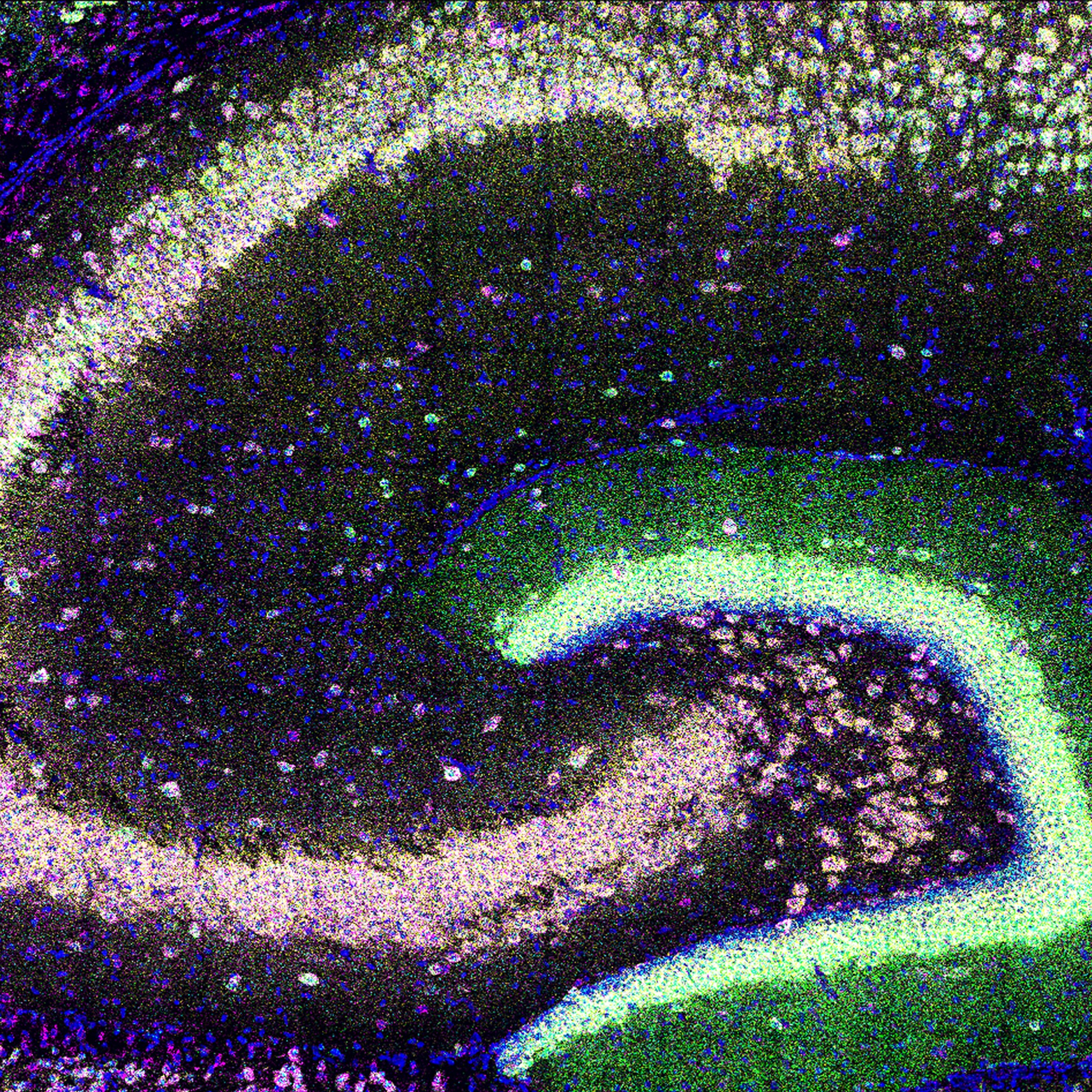
Expression of four neuropil-localized mRNA in hippocampal subregions of a young mouse.
Selective photoactivation and pharmacological manipulations in hippocampal slice cultures reveal a molecular pathway that leads to long-lasting synaptic changes.
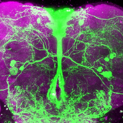
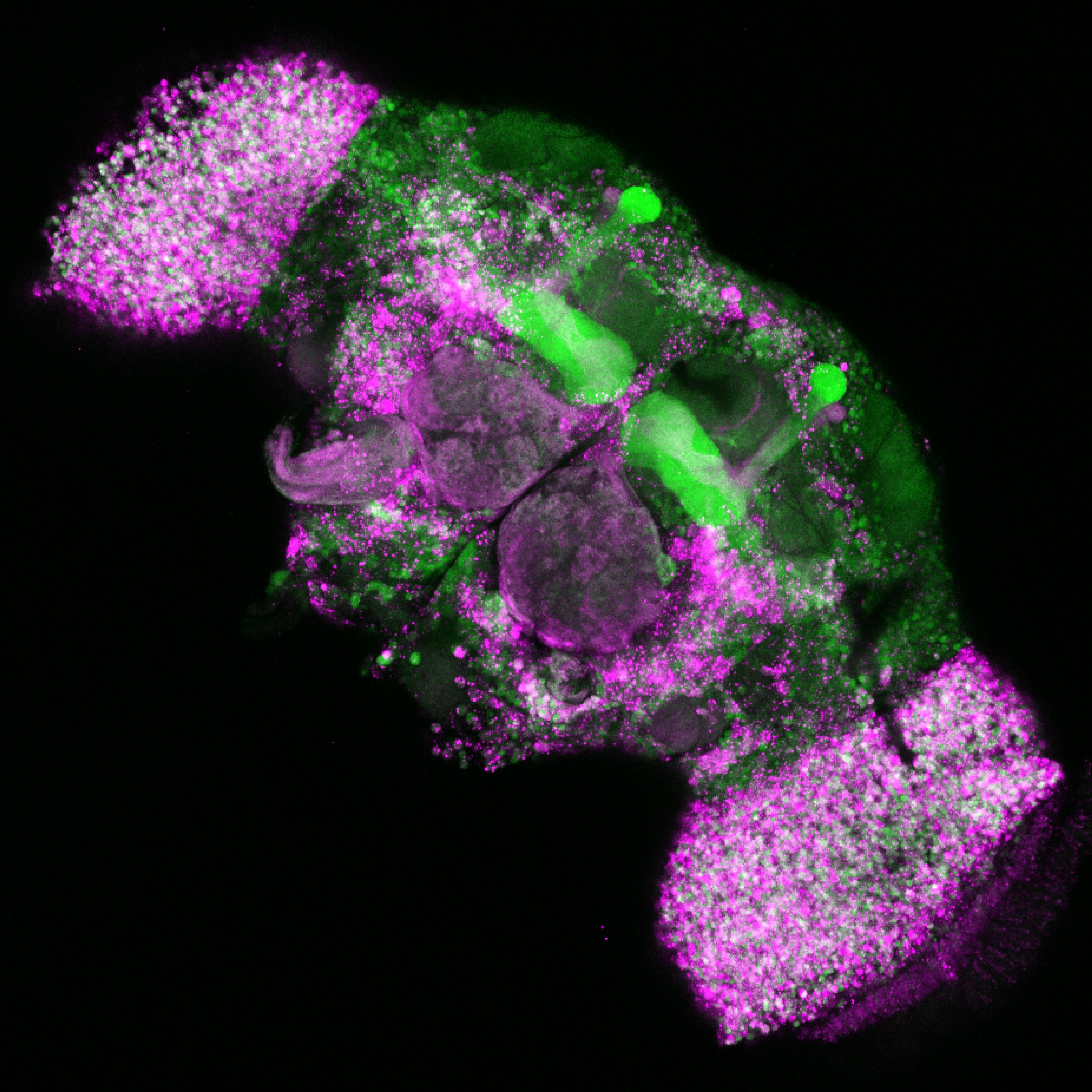
Mapping neuropeptide F receptor expressing-neurons and their role in thirsty water seeking in the adult male fruit fly.
A study on dopamine neurons and fear extinction provides a model for advancing science while being transparent about null results that may not fit the expected narrative.
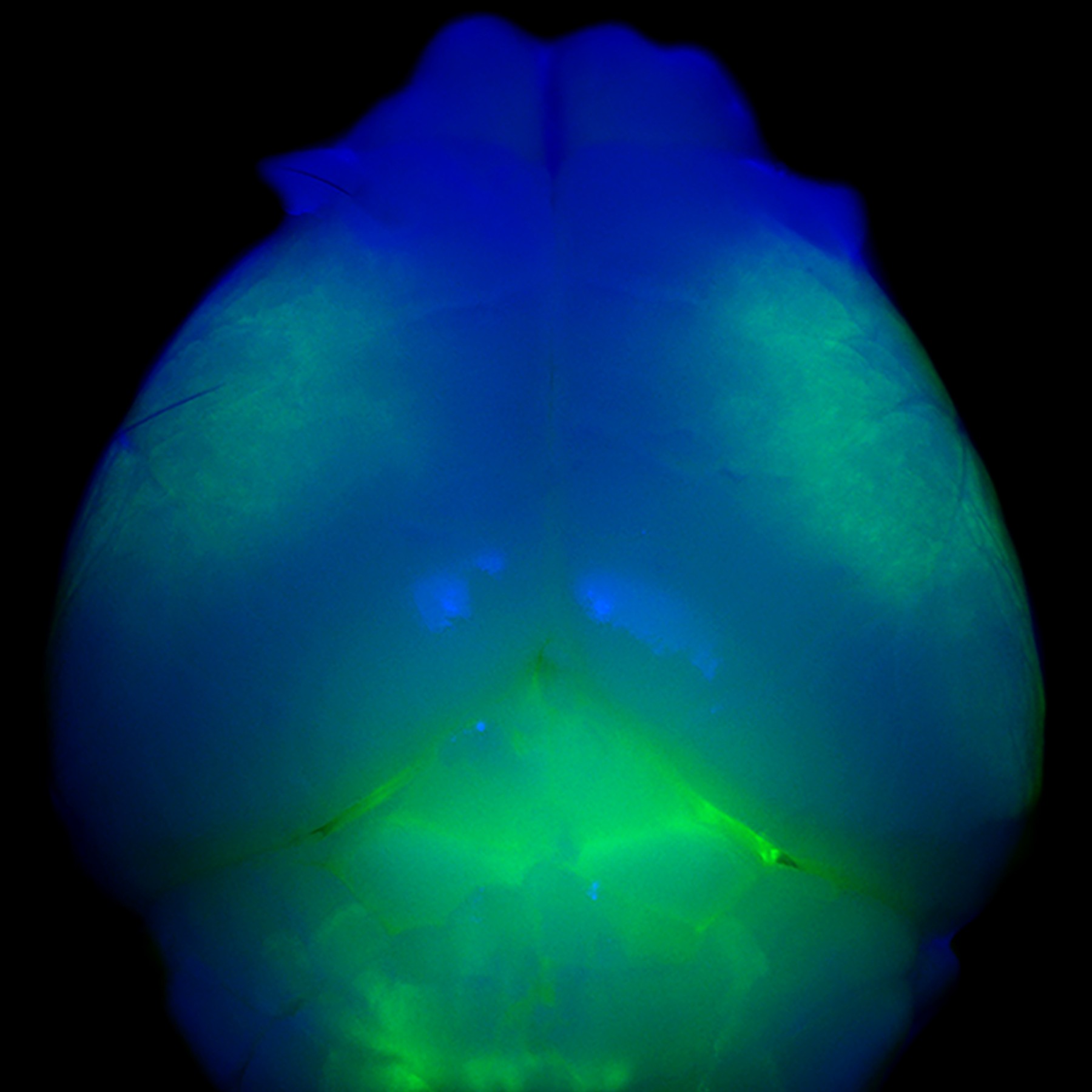
Whole mount view of a young mouse brain with all cortical layer 5 neurons expressing dystonia-related gene Klhl14, highlighted in green.
Exploring how oscillatory and non-oscillatory components of neural activity shape perception and enable multisensory integration and can lead to the McGurk illusion.
Neurons in the adult fruit fly brain have different biases in the splicing of a calcium channel that enables neurotransmission.
Michał Lange entered a neuroscience lab with a strong AI/Computer Science background. Quickly catching up on his biology knowledge, he was able to create a new tool useful to the field.
FOLLOW US
TAGS
CATEGORIES



 RSS Feed


