Snapshots in Neuroscience: Mouse Cerebellum
These images have been selected to showcase the art that neuroscience research can create.
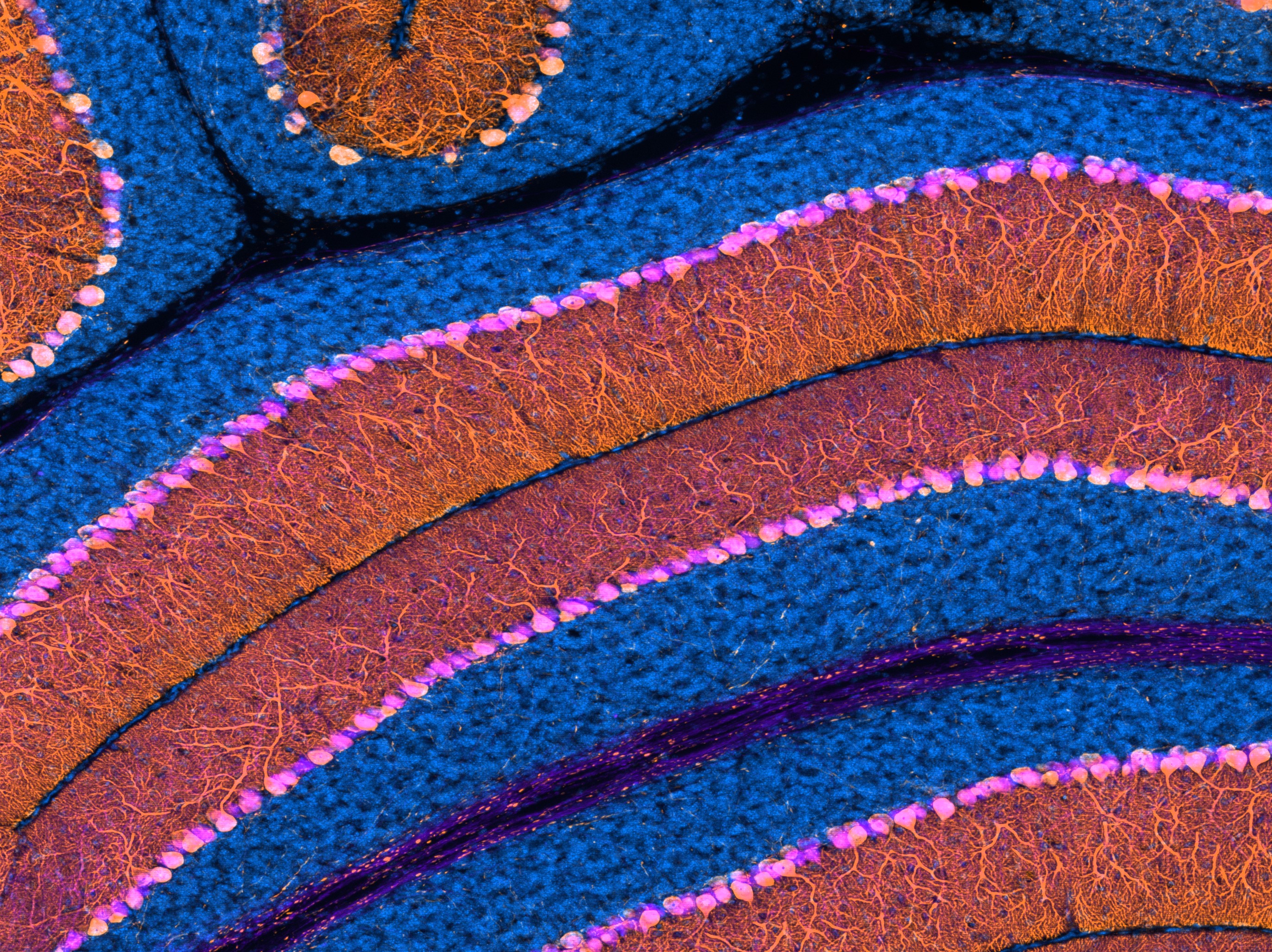
As described by Drs. Jahncke and Wright: Shown is a sagittal section of a mouse cerebellum. The blue channel is a nuclear stain (Hoechst), which is most apparent in granule cells, the most abundant cell type in the brain. The cells labeled with both magenta and orange are Purkinje cells, the primary output cells of the cerebellum. The calcium-binding protein, Calbindin, found on all Purkinje cells, is immunolabelled in magenta. The orange channel is endogenous fluorescence from a Cre-dependent tdTomato reporter driven by Pcp2Cre, which results in Cre expression in all Purkinje cells at the timepoint examined (postnatal day 18).
In our manuscript, we describe the time course and specificity of Cre expression in Pcp2Cre as well as several other Cre lines that drive Cre expression in Purkinje cells and describe how these Cre lines can be used to study synaptic proteins expressed by Purkinje cells.
This 4μm maximum projection image was acquired on a Zeiss Axio Imager M2 fluorescence upright microscope equipped with an Apotome.2 module using a 10X/0.3NA objective.
Read the full article:
Tools for Cre-Mediated Conditional Deletion of Floxed Alleles from Developing Cerebellar Purkinje Cells
Jennifer N. Jahncke and Kevin M. Wright
FOLLOW US
 RSS Feed
POPULAR POSTS
TAGS
CATEGORIES


