Snapshots in Neuroscience: Astrocytes and Neurons
These images have been selected to showcase the art that neuroscience research can create.
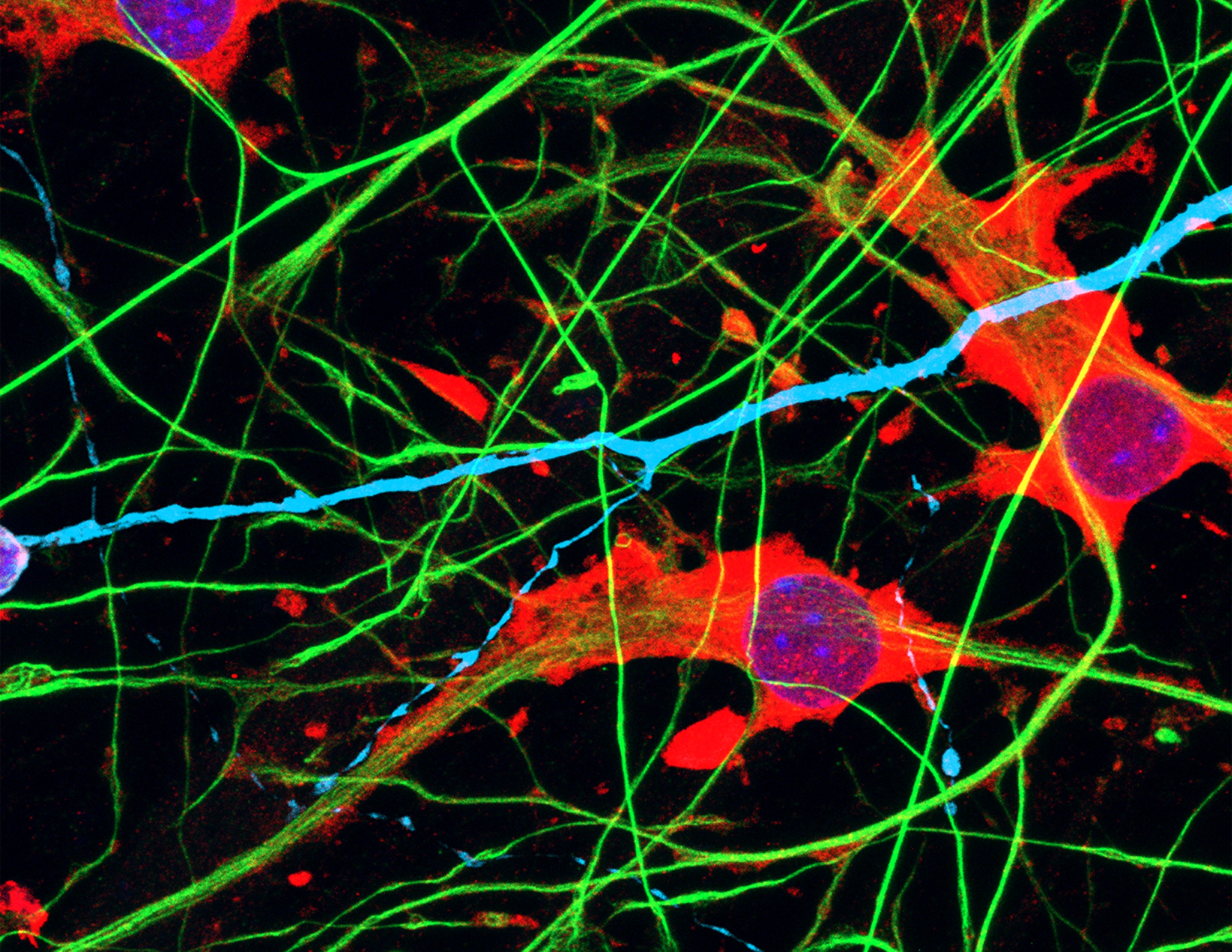
As described by the authors: “This image displays human induced pluripotent stem cell (hiPSC)-derived astrocytes and neurons grown in vitro as a coculture. Astrocytes are labelled using an antibody against the astrocytic markers GFAP (green) and GLAST-1 (red). Neurons are depicted in cyan using an antibody against the neuronal marker MAP2 (cyan). Nuclei are labelled using DAPI (blue).
“In our manuscript, we compare the functionality of hiPSC-derived astrocytes to that of primary rat astrocytes in several assays and in vitro models. Compared to astrocytes in a pure monoculture, astrocytes grown in a coculture with neurons upregulate the expression of the glutamate transporter GLAST-1, a protein involved in the regulation of neurotransmitter concentrations at excitatory glutamatergic synapses. This is one of the findings from our work that supports the bidirectional interaction between neurons and astrocytes.”

Read the full article:
Human Pluripotent Stem Cell-Derived Astrocyte Functionality Compares Favorably with Primary Rat Astrocytes
Bas Lendemeijer, Maurits Unkel, Hilde Smeenk, Britt Mossink, Sara Hijazi, Sara Gordillo-Sampedro, Guy Shpak, Denise E. Slump, Mirjam C.G.N. van den Hout, Wilfred F.J. van IJcken, Eric M.J. Bindels, Witte J.G. Hoogendijk, Nael Nadif Kasri, Femke M.S. de Vrij, and Steven A. Kushner
FOLLOW US
POPULAR POSTS
TAGS
CATEGORIES


 RSS Feed


