Snapshots in Neuroscience: Tracing Cerebellar Afferents
These images have been selected to showcase the art that neuroscience research can create.
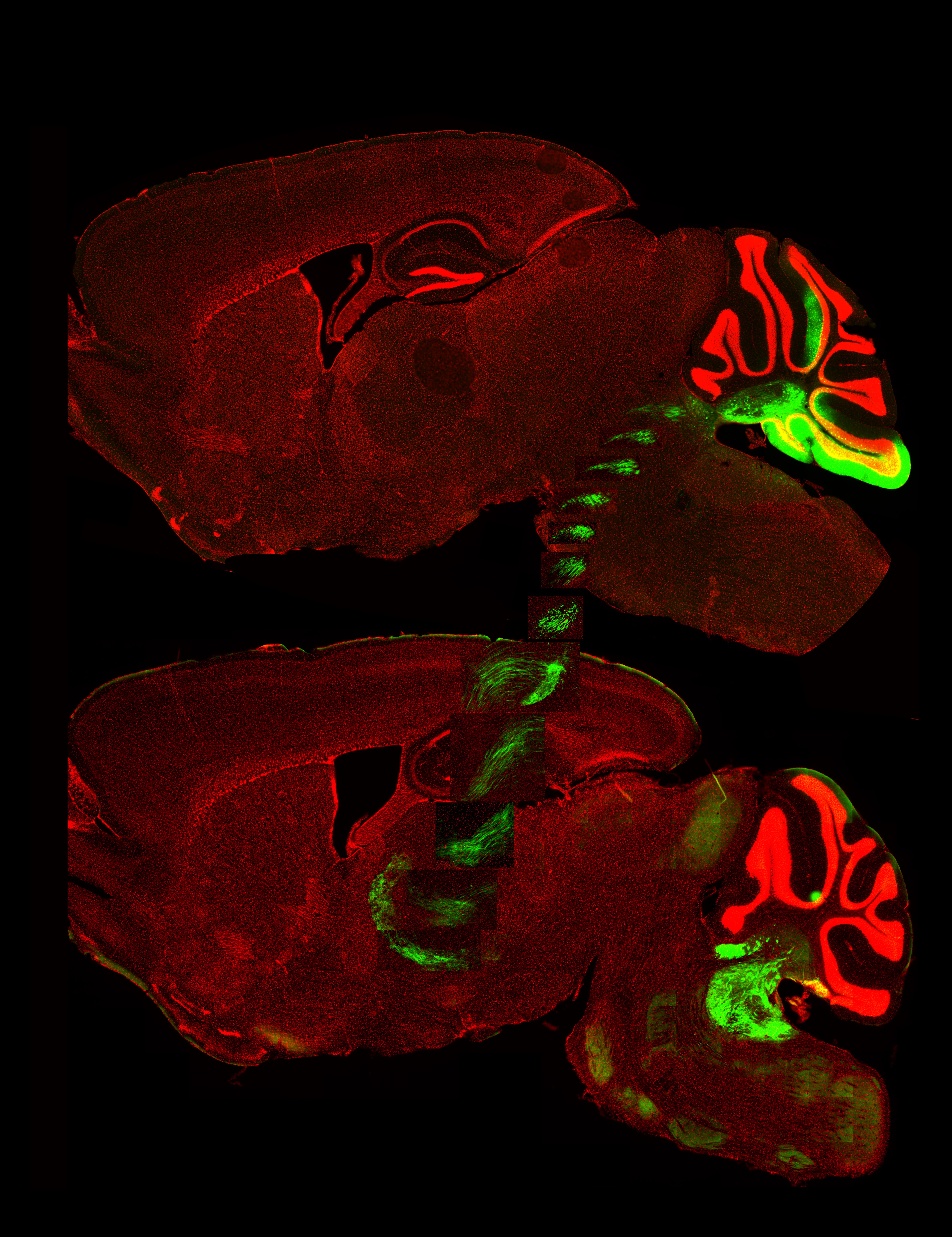
As described by the authors: “Cerebellar nuclei send afferents to the ventral posterolateral nucleus of the thalamus. In the mouse brain these afferents travel over two millimeters through the red nucleus to broadly innervate their destination in the thalamus.
“To collect these images, the mouse cerebellum was transfected with GFP (green) using an AAV1-hSyn-GFP viral vector to label the neurons supplying these afferents. Two weeks following viral injection, the brain was sectioned, nuclei were stained with DAPI (red), and then the slices were imaged using an Olympus VS200 Slide Scanner with a 20x objective.
“The top image is a medial section showing the injection site and the superimposed images show the lateromedial trajectory of the afferents at 200 µm intervals until they reached the thalamus in the bottom section. These images demonstrate the high efficacy of GFP-labeling produced by the AAV1 serotype in the cerebellum, a key finding of our paper, allowing for these long projections to be visualized.”

Read the full article:
Comparative Analysis of Six Adeno-Associated Viral Vector Serotypes in Mouse Inferior Colliculus and Cerebellum
Isabelle Witteveen and Timothy Balmer
FOLLOW US
POPULAR POSTS
TAGS
CATEGORIES


 RSS Feed


