Snapshots in Neuroscience: Corpus Callosum
This image has been selected to showcase the art that neuroscience research can create.
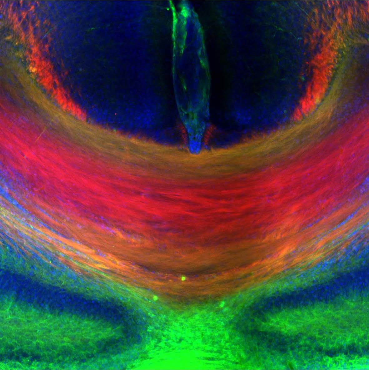
As described by Dr. Martinez-Garay and colleagues: the image shows the midline topographical organization of the corpus callosum of a newborn mouse. L1CAM (green) labels all axons connecting the two cerebral hemispheres, while Neuropilin 1 (red) labels only dorsally located axons. A vibratome 80μm thick coronal section of a mouse brain was stained for L1CAM and Neuropilin 1 and counterstained with DAPI.
The corpus callosum and other main neuronal tracts of the mouse brain were stained as part of a detailed characterization of the PCDH19 knockout mouse, a model for a human encephalopathy. Although the tracts showed no apparent differences between wild type and mutant animals, subtle alterations in neuronal distribution of excitatory and inhibitory neurons were found in the cortex. In addition, mutant animals also displayed behavioral abnormalities.
Remarkably, an effect of animal housing on their subsequent behavior was discovered. Wild type animals exhibited different behavior depending on their cohabitation with mutant animals, an observation with consequences for the selection of control groups.
Read the full article:
Cellular and Behavioral Characterization of Pcdh19 Mutant Mice: subtle Molecular Changes, Increased Exploratory Behavior and an Impact of Social Environment
Natalia Galindo-Riera, Sylvia Adriana Newbold, Monika Sledziowska, Cristina Llinares-Benadero, Jessica Griffiths, Erik Mire, and Isabel Martinez-Garay
FOLLOW US
 RSS Feed
POPULAR POSTS
TAGS
CATEGORIES


