Editor's Pick: What prefrontal cortex?
Laubach and colleagues took a creative approach to address the question "what, if anything, is rodent prefrontal cortex?" posed in the title of their recent article in eNeuro. They carried out a meta-analysis of publications on prefrontal cortex in different species and found marked differences in the kinds of research questions being addressed in mice, rats, monkeys, and humans with regard to prefrontal cortex. To some extent, this is not surprising given the different kinds of research problems that can be optimally addressed in different species, but it also indicates that extrapolation of data obtained in one species to another requires some common anatomical and functional bases for comparison. They also compared anatomical boundaries and criteria for subdivisions within the rodent prefrontal cortex and found inconsistencies in the published literature, in part because different brain atlases use different criteria for delineating the borders of subregions within the frontal cortex. Finally, they carried out a survey of prefrontal cortex researchers and uncovered points of agreement as well as differences in regard to what brain areas constitute "prefrontal cortex" in rodents. Laubach and colleagues propose an anatomical scheme based on morphology of the corpus callosum and position of cortical areas in relation to prefrontal cortex to improve comparisons of prefrontal cortex structure and function across different species.
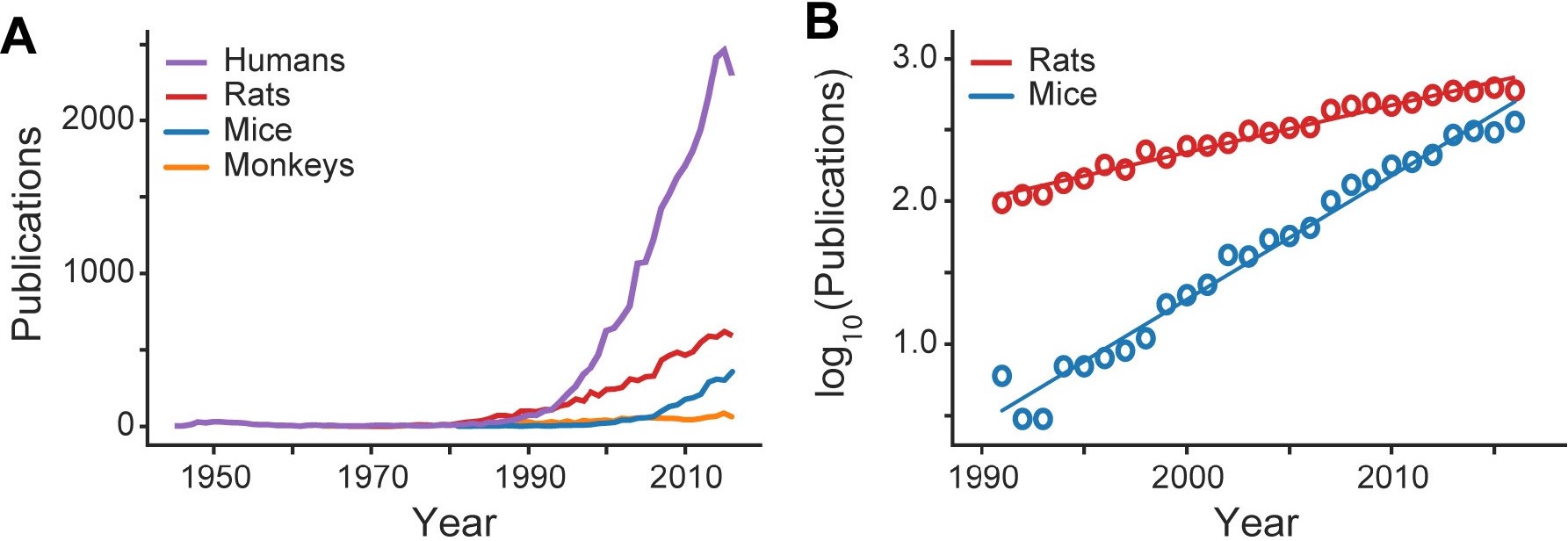
Figure 1: Publication trends in PFC research. A, Publications per year on the PFC of humans, rats, mice, and monkeys from 1945 to 2016. Publication trends on monkey PFC (orange line) have remained unchanged and were not further analyzed. B, Publications on the PFC of mice are appearing at a higher rate than those on rats in the period from 1990 to 2016 (Laubach et al., 2018).
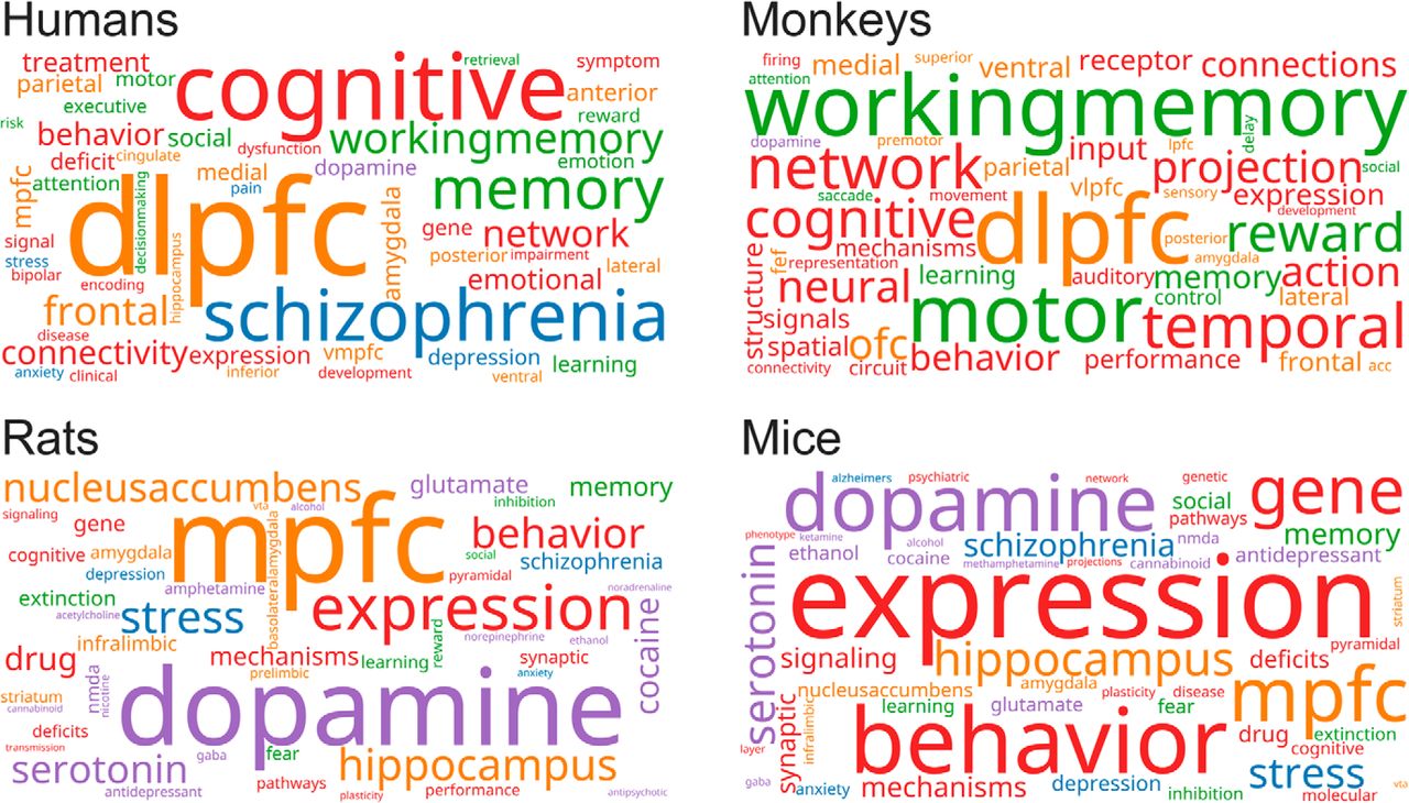
This study is practical about the question it poses. Given the volume of publications on "prefrontal cortex" in mice and rats (Figure 1 of Laubach et al., 2018), it is alarming that there is so much disagreement about which areas comprise the prefrontal cortex in rodents. A common area of controversy arises from asking the question "do rodents have prefrontal cortex?", which focuses on whether they have frontal cortical areas that are anatomically or functionally homologous to specific prefrontal areas in humans and nonhuman primates – typically the dorsolateral prefrontal cortex. But the prefrontal cortex contains many subregions other than dorsolateral, so by applying comparative anatomical criteria to the rodent prefrontal cortex, it is possible to improve the understanding of the functional specialization of the rodent prefrontal cortex and how this might be instructive about human brain function. This framework helps move behavioral and cognitive neuroscience forward by setting the stage for studies in rats and mice using technological approaches uniquely possible in those species, with appropriate constraints on how these studies can be extrapolated to human brain function. This allows experimental work in multiple species to address questions best suited for those species (Figure 2 of Laubach et al., 2018) and avoiding pointless dogma about whether or not rodents have prefrontal cortex at all, or declarations that experiments in one species can fully substitute for experiments in another.
Figure 2: Research focus of prefrontal publications. Word clouds with functional color grouping for papers published on the PFC of mice, rats, monkeys, and humans since 2000. Pharmacological terms are purple. Anatomic terms are orange. Terms associated with diseases and other medical conditions are blue. Psychological constructs are green. Other terms are red (Laubach et al., 2018.)
Read the full article:
What, if anything, is rodent prefrontal cortex?
Mark Laubach, Linda M. Amarante, T. Kyra Swanson and Samantha R. White
FOLLOW US
POPULAR POSTS
TAGS
CATEGORIES


 RSS Feed


