Snapshots in Neuroscience: Synaptic Boutons
This image has been selected to showcase the art that neuroscience research can create.
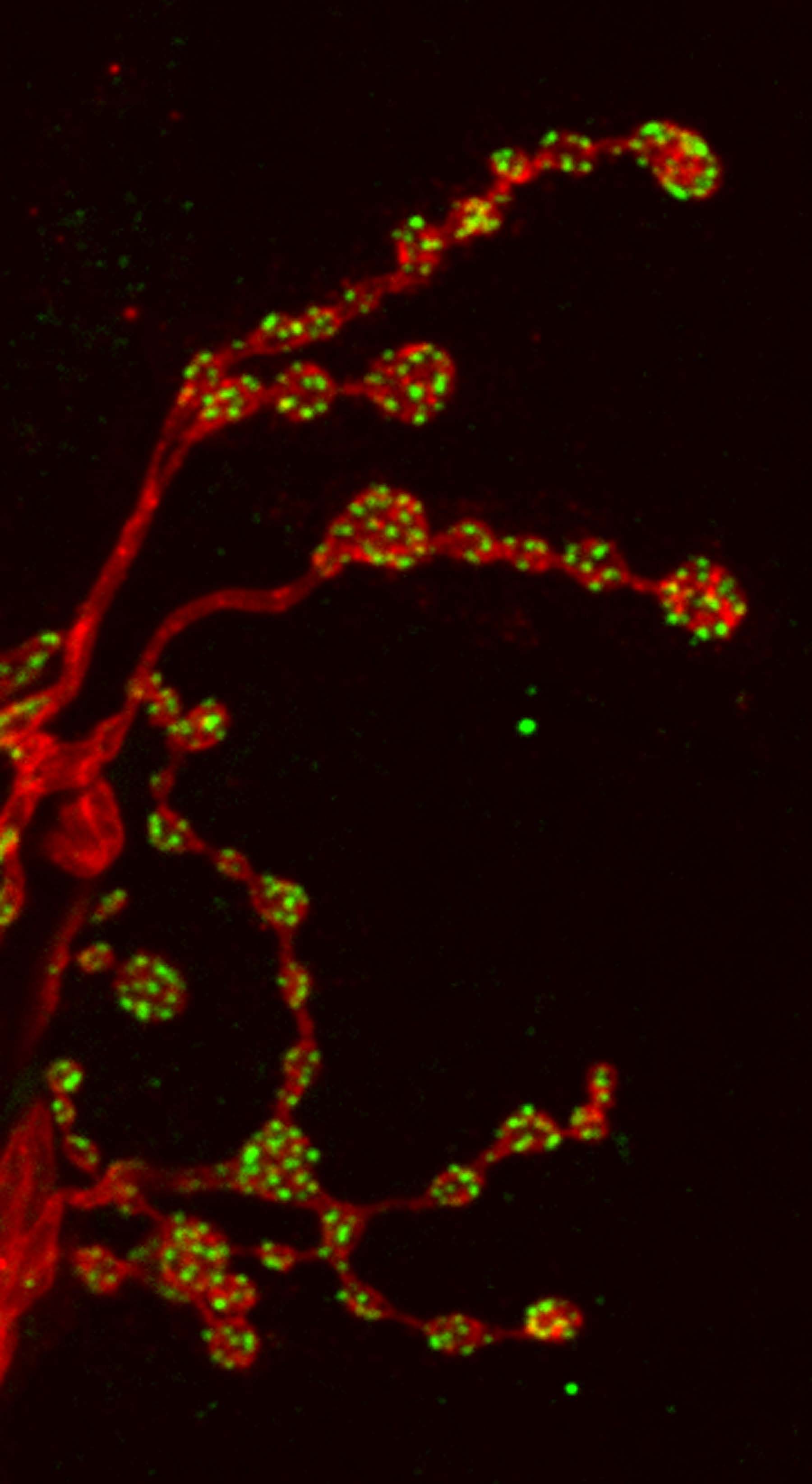
As described by Dr. van Swinderen and colleagues: The muscles of the larval fruit fly are innervated by two types of motoneurons: 1b and 1s. The synaptic boutons of these neurons (shown in red) can be seen in the image. 1b boutons are larger and have more release sites (shown in green) while 1s boutons are smaller and have fewer release sites. This study found that not all release sites are active in 1b boutons, whereas most are active in the smaller 1s boutons.
The Drosophila neuromuscular junction can be used to study the effects of general anaesthetics on synaptic release. The intravenous anaesthetic propofol decreases the number of active sites proportionally by half in 1b and 1s boutons, without affecting release probability of remaining active sites.
Read the full article:
Proportional Downscaling of Glutamatergic Release Sites by the General Anesthetic Propofol at Drosophila Motor Nerve Terminals
Shanker Karunanithi, Drew Cylinder, Deniz Ertekin, Oressia H. Zalucki, Leo Marin, Nickolas A. Lavidis, Harold L. Atwood, and Bruno van Swinderen
FOLLOW US
 RSS Feed
POPULAR POSTS
TAGS
CATEGORIES


