Snapshots in Neuroscience: Sensory Neurons
June 25, 2020
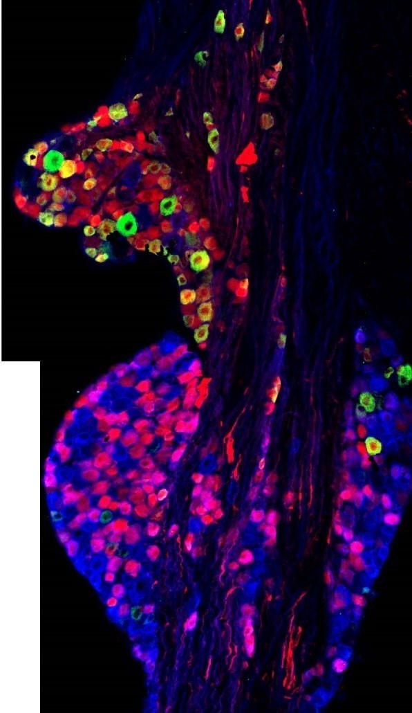
This image has been selected to showcase the art that neuroscience research can create.
As described by Dr. Taylor-Clark and colleagues: This confocal 20X image shows the neuronal cell bodies of sensory nerves within the vagal ganglia, just outside the brainstem. These neurons (~15-35um in diameter) project throughout the visceral organs, providing sensory nerve endings that sense the local environment. These nerves are heterogeneous with respect to protein expression, structure and function. A subset are activated by noxious stimuli and these express the receptor for chili (TRPV1), which are identified here in red (TRPV1-tdTomato mouse).
Vagal ganglia are comprised of 2 smaller ganglia which express different proteins. Here jugular ganglion neurons at the top are identified by the green staining (antibody against neurotrophin receptor TRKA), whereas the nodose ganglion neurons are identified by the blue staining (antibody against neurotrophin receptor TRKB). Overall, this image shows the substantial heterogeneity of vagal sensory neurons, consistent with these divergent roles in autonomic regulation of multiple organs.
Read the full article:
Mapping of Sensory Nerve Subsets within the Vagal Ganglia and the Brainstem Using Reporter Mice for Pirt, TRPV1, 5-HT3, and Tac1 Expression
Seol-Hee Kim, Stephen H. Hadley, Mikayla Maddison, Mayur Patil, Byeong Cha, Marian Kollarik, and Thomas E. Taylor-Clark
Category: Snapshots in Neuroscience
FOLLOW US
 RSS Feed
POPULAR POSTS
TAGS
CATEGORIES


