Snapshots in Neuroscience: Fusiform Cell
March 18, 2021
This image has been selected to showcase the art that neuroscience research can create.
As described by Dr. Thawann Malfatti and colleagues: This is a confocal image showing a neuron, likely a fusiform cell, at the dorsal cochlear nucleus (DCN) of a coronal slice from a mouse expressing CaMKIIα-ChR2-eYFP along its somatic membrane and dendrites. DAPI staining of cell nuclei is shown in blue.This particular cryostat 30µm-thick section shows that, following injection of a rAAV5/CamK2-hChR(H134R)-eYFP viral vector, the neuron can express and place ChR2-eYFP proteins in the membrane, allowing it to be optogenetically activated. Because only a subpopulation of neurons in the DCN express the CaMKIIα protein, this procedure allows for targeting those neurons for artificial controlling or monitoring nerve cell activity.
Using this method, we found that: neurons of different morphology are targeted, optogenetic modulation of those neurons changed the circuitry activity, and concomitant sound stimulation interferes with the optogenetically-driven changes.
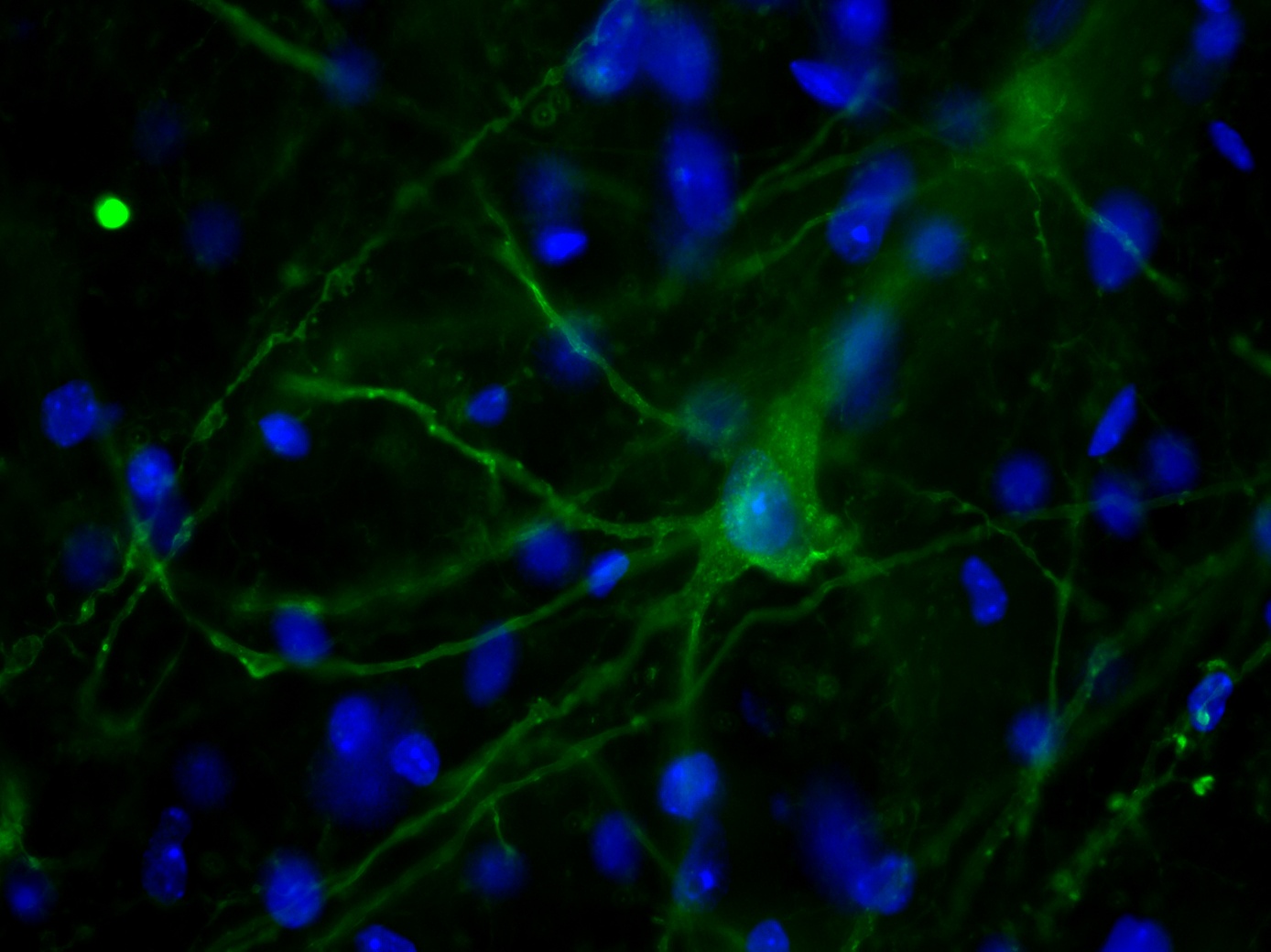 This image was captured with a Zeiss Examiner Z1 confocal microscope, using a Plan-Neochromat 40x/0.75 objective.
Read the full article:
Using Cortical Neuron Markers to Target Cells in the Dorsal Cochlear Nucleus
Thawann Malfatti, Barbara Ciralli, Markus M. Hilscher, Steven J. Edwards, Klas Kullander, Richardson N. Leao and Katarina E. Leao
Category: Snapshots in Neuroscience
FOLLOW US
 RSS Feed
POPULAR POSTS
TAGS
CATEGORIES


