Snapshots in Neuroscience: Cortical Neurons
June 10, 2021
This image has been selected to showcase the art that neuroscience research can create.
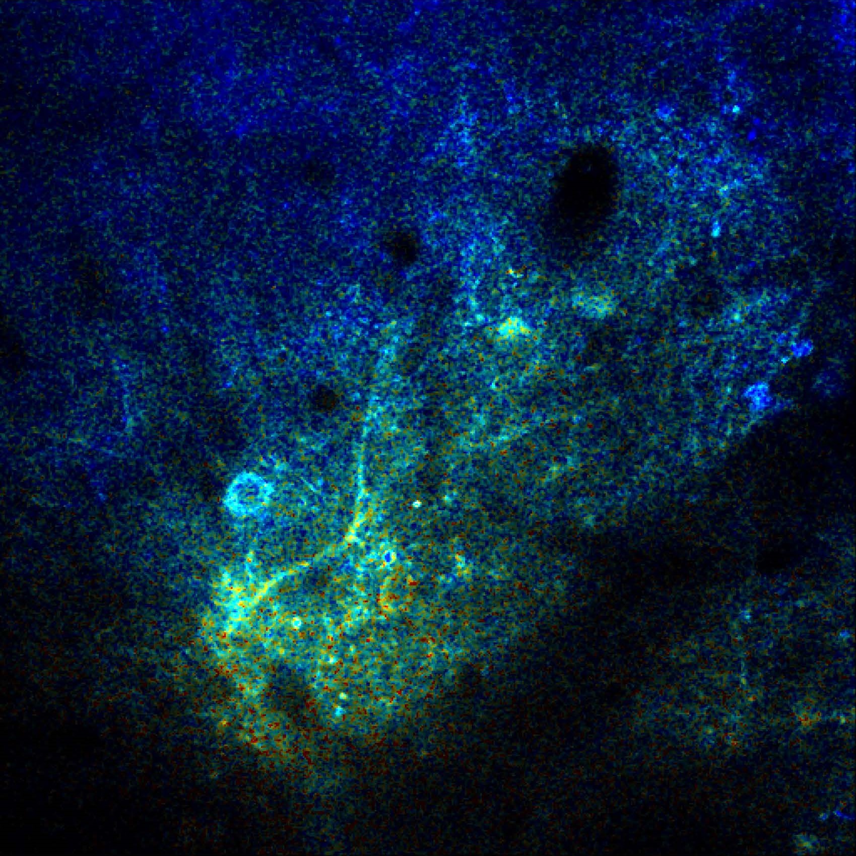
As described by Dr. Ksenia Kastanenka and colleagues: This is an image of cortical neurons in a mouse model of Alzheimer's disease taken with high-resolution multi-photon microscopy through a cranial window.The neurons express a genetically encoded ratiometric calcium indicator, YellowCameleon 3.6, that allows determination of absolute calcium levels. Normally cortical neurons keep intracellular calcium within a tight range, around 100nM (see blue processes and cell bodies). However, amyloid beta present in the brains of these mice trigger calcium elevations within neurons (see orange and yellow processes as well as cell bodies).
A novel botanical drug NB-02 restored neuronal calcium homeostasis. Thus NB-02 is a promising potential therapy for Alzheimer's disease.
Read the full article:
Novel Botanical Therapeutic NB-02 Effectively Treats Alzheimer’s Neuropathophysiology in an APP/PS1 Mouse Model
Yee Fun Lee, Lavender Lariviere, Alyssa N. Russ, Sang-Zin Choi, Brian J. Bacskai and Ksenia V. Kastanenka
Category: Snapshots in Neuroscience
FOLLOW US
 RSS Feed
POPULAR POSTS
TAGS
CATEGORIES


