Snapshots in Neuroscience: Hippocampus
This image has been selected to showcase the art that neuroscience research can create.
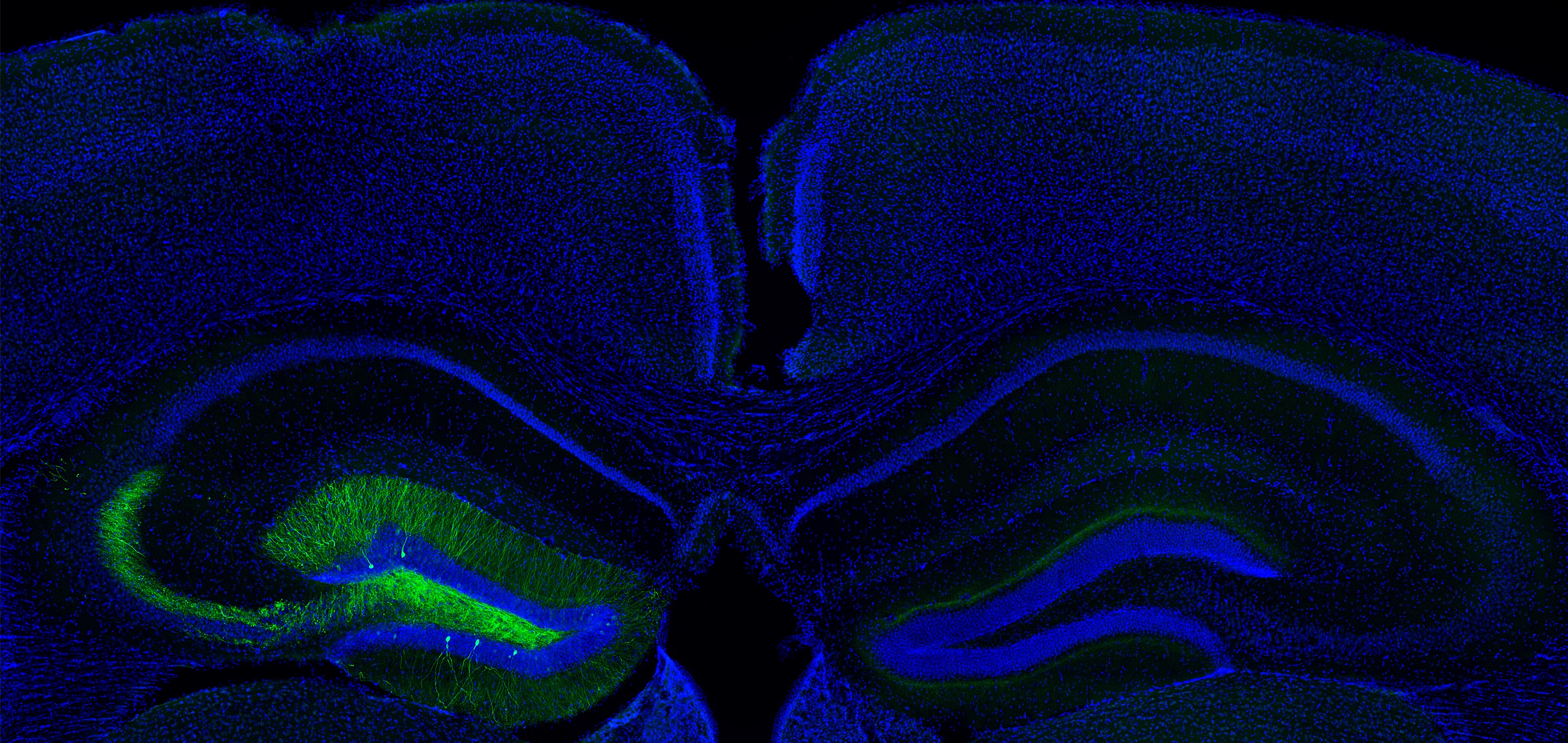
As described by Dr. Maithe Arruda-Carvalho and colleagues: The photo shows a section of a mouse brain hippocampus, a region that is widely studied in neuroscience because of its role in learning and memory. A cre-dependent adeno-associated virus encoding green fluorescent protein (GFP) was injected into the dentate gyrus of the left hippocampus of a wild-type mouse and visualized with an antibody against GFP. The blue signal is a nuclear stain (Hoechst 33342) which is often used to identify brain regions and their subfields.
Normally, one would expect to see minimal GFP expression in a wild-type mouse because the virus is engineered only to express GFP in transgenic mice that express Cre (which wild-type mice lack). However, amplification of the GFP using antibodies revealed notable off-target expression in mice that are often designated as controls.
These findings suggest that there can be significant Cre-independent expression in subjects typically designated as controls and suggest that researchers must cautiously design and interpret experiments that use Cre-dependent AAV infection.
Read the full article:
Off-Target Expression of Cre-Dependent Adeno-Associated Viruses in Wild-Type C57BL/6J Mice
Justin J. Botterill, Abdessattar Khlaifia, Brandon J. Walters, Mark A. Brimble, Helen E. Scharfman, and Maithe Arruda-Carvalho
FOLLOW US
 RSS Feed
POPULAR POSTS
TAGS
CATEGORIES


