Snapshots in Neuroscience: Cerebellar Cortex
This image has been selected to showcase the art that neuroscience research can create.
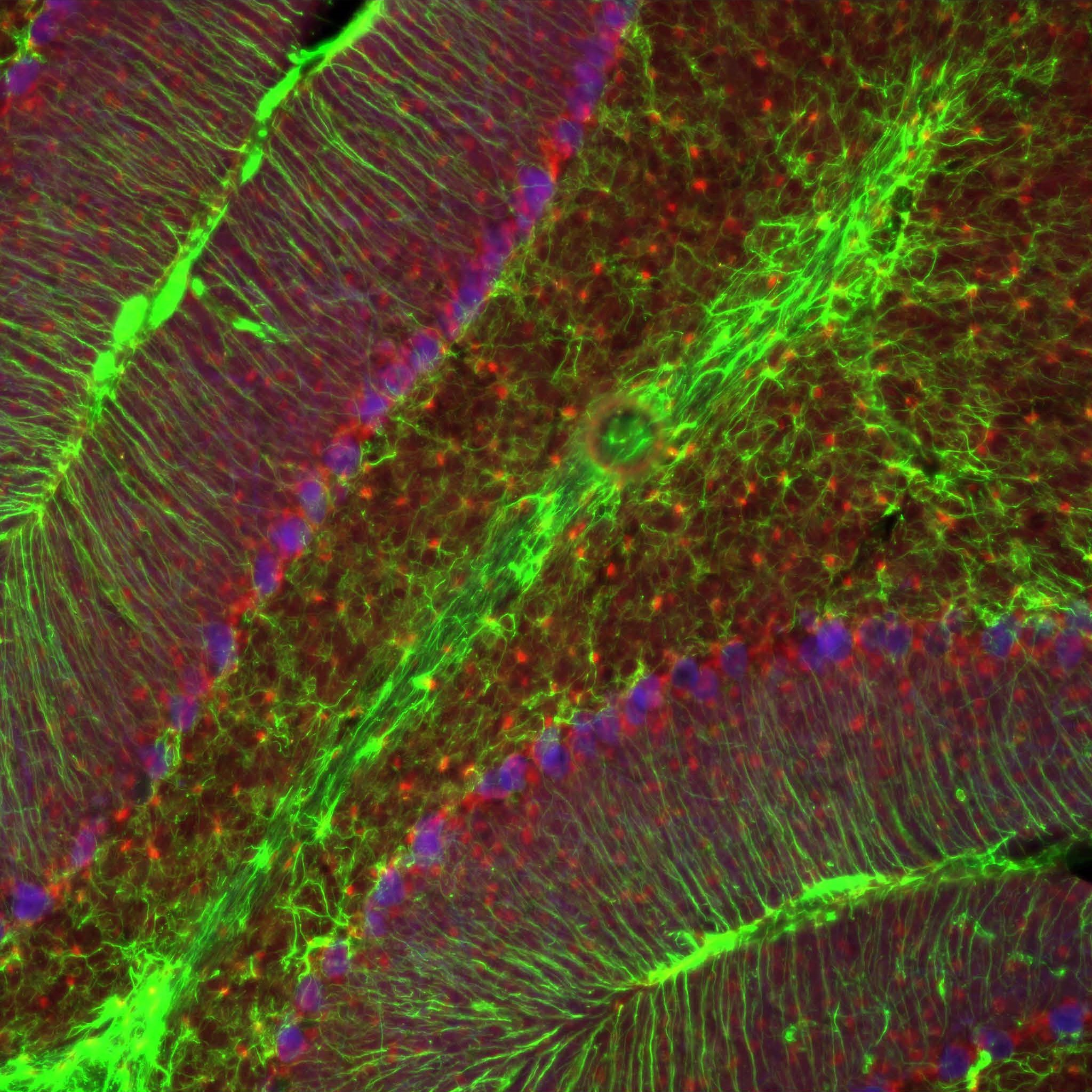
As described by Dr. Gebril and colleagues: The image shows part of the juvenile cerebellar cortex of a mouse at postnatal day 21. This is a triple immunofluorescence staining for glial fibrillary acidic protein (GFAP, green), calbindin (blue) and adenosine kinase (ADK, red).
This image was taken within the context to analyze the expression of ADK during cerebellar development. ADK, the main regulator of adenosine, undergoes coordinated expression changes during brain development and its isoform ADK-L, expressed in the cell nucleus, plays a role in neurogenesis and cell proliferation.
Here we show that ADK expression is associated with the proliferation of young neurons and with development of the cerebellum in mice.
Read the full article:
Developmental Role of Adenosine Kinase in the Cerebellum
Hoda Gebril, Amir Wahba, Xiaofeng Zhou, Tho Lai, Enmar Alharfoush, Emanuel DiCicco-Bloom, and Detlev Boison
FOLLOW US
 RSS Feed
POPULAR POSTS
TAGS
CATEGORIES


