Snapshots in Neuroscience: Spatial Memory-Activated Neurons
This image has been selected to showcase the art that neuroscience research can create.
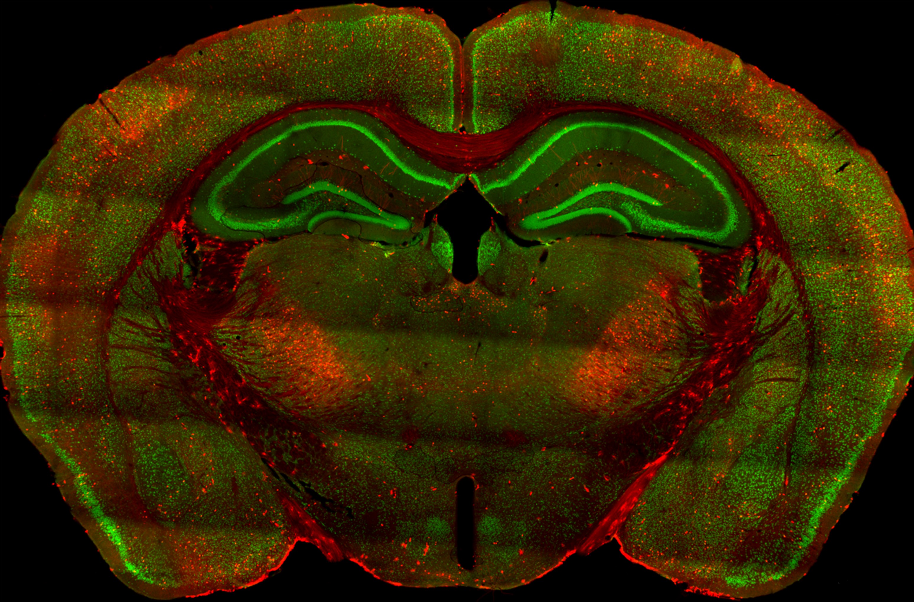
As described by Naik, Brodovskaya and colleagues: The following image is a coronal brain slice of activity reporter TRAP2 mice that labels, with tdTomato (red), neuronal ensembles active during a rewarded T-maze spatial memory task. Mice acquired the rewarded alteration memory on the second day of training because they demonstrated improved performance on the subsequent days.
The confocal image (10x) is at the level of the hippocampus, where tdTomato (red) labels spatial memory-activated neurons, and NeuN (green) labels neurons. Spatial memory-activated neurons were present in the retrosplenial cortex, hippocampus (CA1, DG), and mediodorsal thalamic nucleus.
A seizure prevented the recall of alteration memory and also activated memory-labeled structures. There was a widespread overlap between learning-activated ensembles and seizure-activated neurons. We propose that seizures cause retrograde amnesia by engaging the circuits that participate in memory consolidation.
Read the full article:
Extrahippocampal Seizure and Memory Circuits Overlap
Aijaz Ahmad Naik, Anastasia Brodovskaya, Smriti Subedi, Amman Akram, and Jaideep Kapur
FOLLOW US
 RSS Feed
POPULAR POSTS
TAGS
CATEGORIES


