Snapshots in Neuroscience: Developing Vagal Sensory Cell Types
This image has been selected to showcase the art that neuroscience research can create.
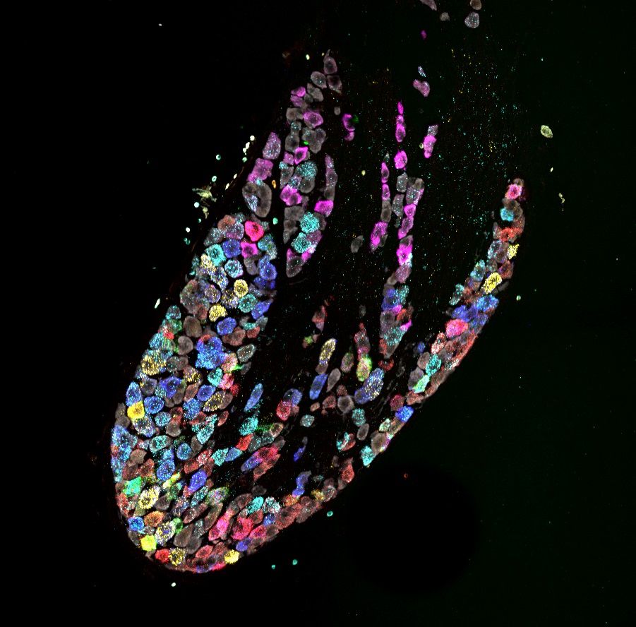
As described by Dr. Kamitakahara and colleagues: Modality specific sensory neuron types are required for detection of different gastrointestinal stimuli, such as gastrointestinal stretch or ingested nutrients. These different sensory cell types can be identified by gene expression markers.
Using in situ hybridization for cell type specific marker genes, this sample from postnatal day (P) 7 mouse nodose ganglion illustrates the developmental emergence of identifiable vagal sensory cell types. For example, neurons expressing Calca (magenta) that project to the stomach are distinct from neurons expressing Vip (yellow) that project to the intestine. In our study, we demonstrate that distinct cell type specific gene expression patterns are not well defined prenatally but emerge within the first postnatal week in mice as they begin to ingest milk.
This image was captured using confocal microscopy at 40x magnification in 20μm tissue sections from the nodose ganglion. It is the composite of 5 images captured following each of 5 rounds of labeling and reprobing for a total of 12 mRNA transcripts and neuronal marker (HuCD protein; gray).
Read the full article:
Ontogeny and Trophic Factor Sensitivity of Gastrointestinal Projecting Vagal Sensory Cell Types
Meaghan E. McCoy and Anna K. Kamitakahara
FOLLOW US
 RSS Feed
POPULAR POSTS
TAGS
CATEGORIES


