Snapshot in Neuroscience: Reconstructing Photoreceptor Terminals
This image has been selected to showcase the art that neuroscience research can create.
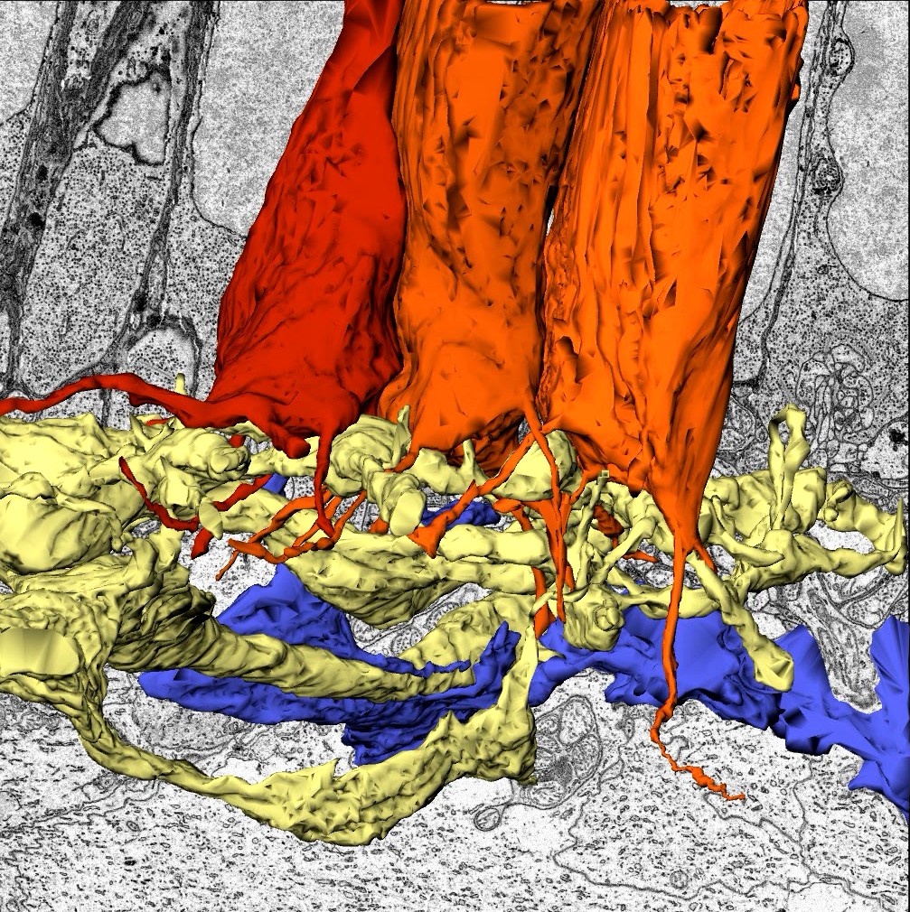
As described by Dr. Anastassov and colleagues: this image is from the retina of the cartilaginous fish Little skate — a vertebrate with only one type of photoreceptor cell. Skate photoreceptors are unusual in their ability to retain function over a full range of illumination, a task usually delegated to multiple photoreceptor cell types like rods and cones. The image shows 3D reconstructions of skate photoreceptor synaptic terminals (shades of orange) and some of their selected postsynaptic partners (yellow and blue). Data was obtained from serial block-face electron microscopy performed on a small region in the outer plexiform layer covering a volume of ~1.65x104µm3. In the background, one of the raw electron microscopy images in this volume can be seen with the nuclei, synaptic vesicles and synaptic ribbons of photoreceptors distinguishable.
Skate photoreceptors exhibit unusual structural characteristics that are either common to rod and cone photoreceptors in other vertebrates or are somewhere in between those of typical vertebrate rods or cones. For example, the number of synaptic ribbons in different terminals varies because multiple postsynaptic partners make invaginating connections to each photoreceptor terminal, and there are long extending telodendria that connect neighboring photoreceptors.
Magaña-Hernández et al. performed serial block-face 3D electron microscopy imaging (SB-3DEM) on epoxy resin-embedded tissue stained with uranyl acetate. The dataset used and analyzed in this image (P2R9 volume) is from a region of interest in the outer plexiform layer with a width and height of 27.6μm and a depth of 21.5μm; voxel size was 4.5x4.5x70nm (xyz). A Zeiss Sigma VP system, equipped with a Gatan 3View in-chamber ultramicrotome stage, was used for imaging.
Read the full article:
Ultrastructural characteristics and synaptic connectivity of photoreceptors in the simplex retina of Little skate (Leucoraja erinacea)
Laura Magaña-Hernández, Abhiniti S. Wagh, Jessamyn G. Fathi, Julio E. Robles, Beatriz Rubio, Yaqoub Yusuf, Erin E. Rose, Daniel E. Brown, Priscilla E. Perry, Elizabeth Hamada, and Ivan A. Anastassov
FOLLOW US
 RSS Feed
POPULAR POSTS
TAGS
CATEGORIES


