Snapshots in Neuroscience: Hippocampal Somatostatin Interneurons
This image has been selected to showcase the art that neuroscience research can create.
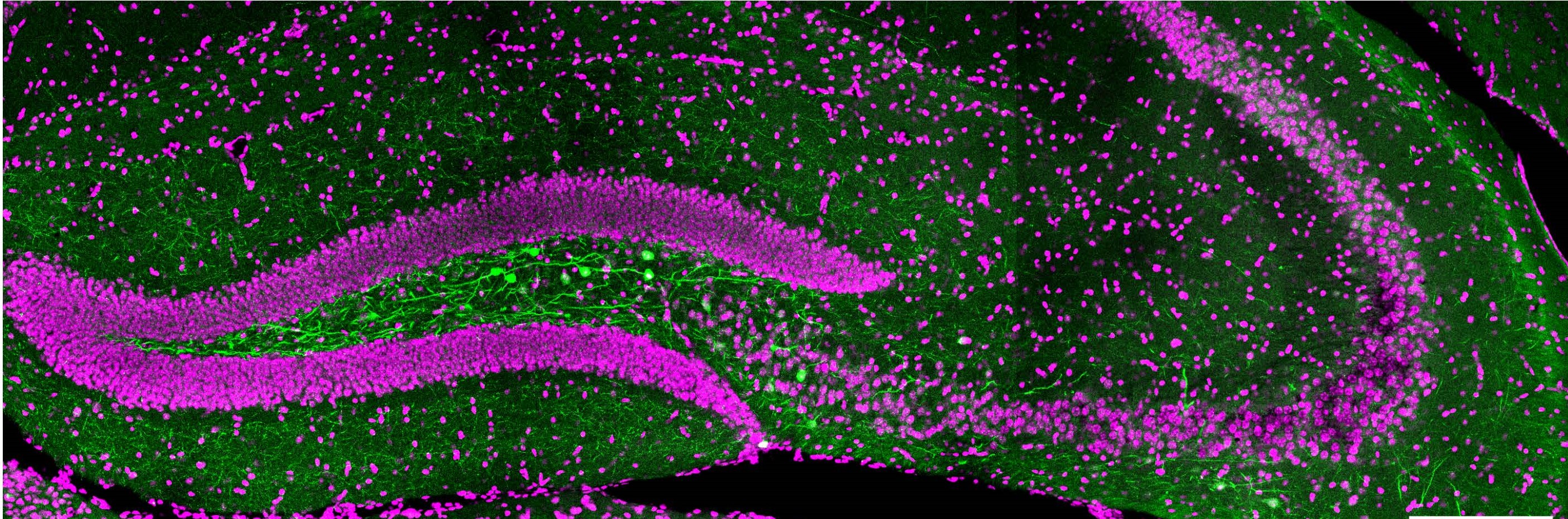
As described by Dr Méndez and colleagues: This confocal microscopy image shows a coronal section of the hippocampus of a mouse. The nuclei of all brain cells are shown in magenta using DAPI, a fluorescent dye that binds the nuclear DNA. In green is a population of inhibitory neurons that express the marker somatostatin (SST).
The SST inhibitory neurons express a Green Fluorescent Protein coupled to the optogenetic actuator ChETA, which Hernandez-Vivanco et al. used in their experiments to activate SST neurons with blue light. The green fluorescent protein allows visualization of SST-expressing inhibitory neurons extending their dendrites and axons locally in the hippocampus, to contact and be contacted by neighboring cells.
However, some SST inhibitory neurons send axon branches outside the hippocampus, contacting neurons located in a distal brain region. These axons form inhibitory synaptic contacts with medial septum neurons and have an important role in synchronizing the activity of these two distant brain regions involved in memory formation and recall.
Read the full article:
Protein kinase A-dependent plasticity of local inhibitory synapses from hilar somatostatin-expressing neurons.
Alicia Hernandez-Vivanco, Esther Jimenez-Redondo, Nuria Cano-Adamuz, and Pablo Mendez
FOLLOW US
 RSS Feed
POPULAR POSTS
TAGS
CATEGORIES


