Snapshots in Neuroscience: Mouse Retina
This image has been selected to showcase the art that neuroscience research can create.
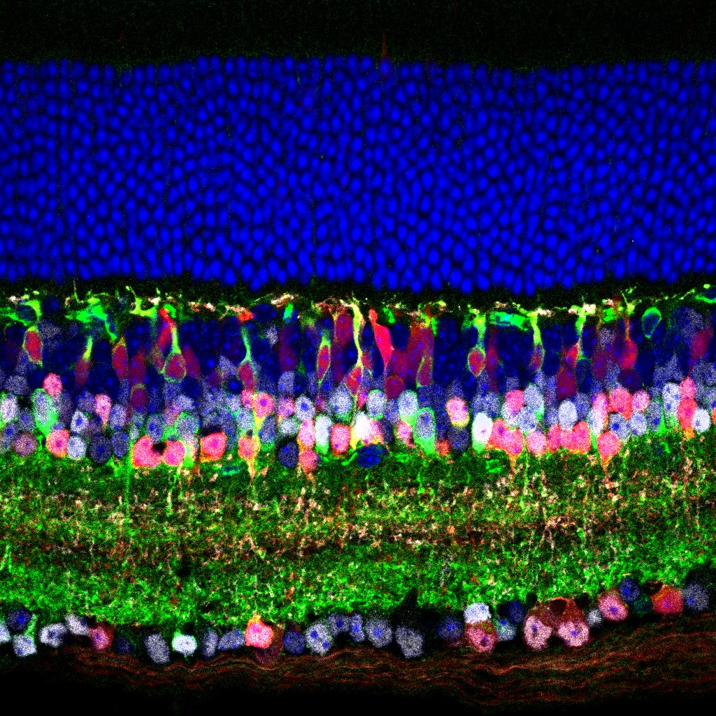
As described by Drs. Zoghbi and Wu: this image shows the laminar structure of the adult mouse retina. The retina is the light-sensitive tissue lining the back of the eye and is critical for visual perception. Zoghbi and Wu crossed an Atoh1-Cre knock-in mouse model to a Cre-dependent TdTomato reporter line and observed a subset of TdTomato-labeled cells (red) in the mouse retina. They performed immunostaining using a panel of cell type-specific markers to characterize the identity of the TdTomato-labeled cells.
In this image, a 10µm-thick section of mouse retina was stained for Pax6 (gray) and PKARIIβ (green), markers for amacrine cells and type 3b bipolar cells, respectively, and DAPI (blue) for nuclei. The section was imaged with 20X objective by Zeiss LSM710 confocal microscope. The partial overlap between the TdTomato and Pax6 or PKARIIβ suggests the Atoh1-Cre knock-in mouse model ectopically expresses Cre in the retina and selectively labels a subset of amacrine cells and bipolar cells.
Read the full article:
The Atoh1-Cre Knock-In Allele Ectopically Labels a Subpopulation of Amacrine Cells and Bipolar Cells in Mouse Retina
Sih-Rong Wu and Huda Y. Zoghbi
FOLLOW US
 RSS Feed
POPULAR POSTS
TAGS
CATEGORIES


