Snapshots in Neuroscience: In vivo and ex vivo cross-section images of tree shrew retinas
These images have been selected to showcase the art that neuroscience research can create.
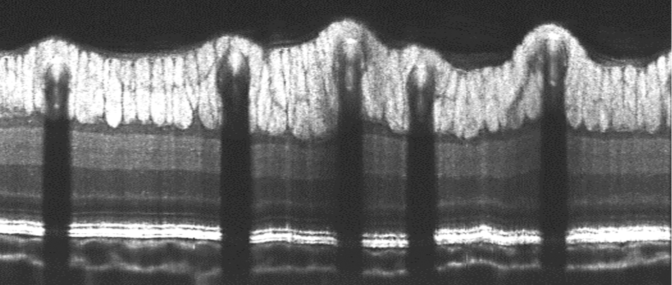
As described by Dr. Liu and colleagues: Below are cross-section images of tree shrew retinas near the optic nerve head (ONH) region.
The in vivo visible-light optical coherence tomography (vis-OCT) B-scan image below shows vertically elongated and densely packed axon bundles in the retinal nerve fiber layer (RNFL), a distinct dark band of the ganglion cell layer (GCL), a clear sub-layered inner plexiform layer (IPL), and a dark band of the inner nuclear layer (INL).
For in vivo imaging, tree shrews were anesthetized using isoflurane with supplemental oxygen, followed by an intraperitoneal cocktail injection of ketamine and xylazine. This image was acquired with a small animal vis-OCT system, Halo 100 Opticent Health, capturing a total volume of 1.12 mm × 1.5 mm (x × y) with an axial resolution of 1.46 μm/pixel and a lateral resolution of 2.18 μm/pixel.
The retinal layer organization detected in vivo by the vis-OCT technique was validated with corresponding ex vivo confocal images of retinal sections immunostained with different retinal markers.
The ex vivo confocal image below is one such image. The retina cross section was stained with Tuj-1 to highlight axons (in green) of retinal ganglion cells (RGCs), Glial Fibrillary Acidic Protein (GFAP) to mark astrocytes and Müller glial cells (in red), and DAPI (4′,6-diamidino-2-phenylindole) for cell nuclei (in blue). This image highlights vertically elongated axon bundles (in green) tightly wrapped by astrocytes (in red) on the top surface, with 3-5 layers of nuclei in the ganglion cell layer (GCL) visible beneath.
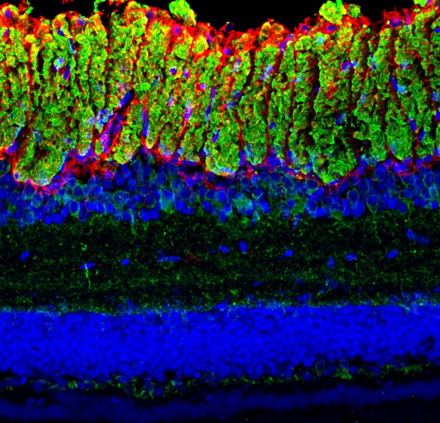
This staining technique confirmed the organization of axon bundles as detected in vivo. In the central area of the tree shrew retina, the axon bundles appeared to be densely packed, vertically elongated, and stratified, resembling human eyes more closely than mouse eyes.
To capture this image, tree shrew eyes were dissected and fixed in PFA for 30 minutes, followed by cryoprotection in 30% sucrose overnight. The eyecups were sectioned at 20-30 μm using a cryostat. Sections containing the ONH were used for this study. Primary antibodies included Tuj-1 (a gift from Tony Spano at UVA, 1:200) and GFAP (Abcam #ab4674, 1:200). This confocal image was captured using the 3D Z-stack mode on a Zeiss microscope (LSM 800, Carl Zeiss) with a magnification of 20× and 0.64 μm/pixel resolution
Read the full article:
Comparative In Vivo Imaging of Retinal Structures in Tree Shrews, Humans, and Mice
Marta Grannonico, David A. Miller, Mingna Liu, Michael A. Krause, Elise Savier, Alev Erisir, Peter A. Netland, Jianhua Cang, Hao F. Zhang, and Xiaorong Liu
FOLLOW US
 RSS Feed
POPULAR POSTS
TAGS
CATEGORIES


