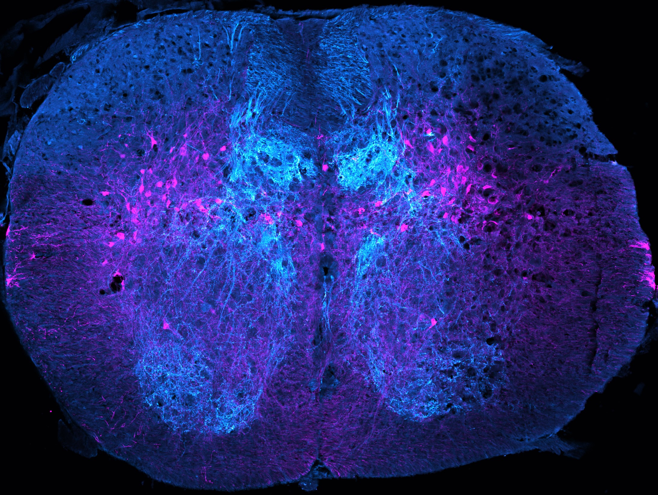
Snapshots in Neuroscience: Mouse spinal cord
These images have been selected to showcase the art that neuroscience research can create.
As described by Dr. Helen Lai and colleagues: This image depicts a transverse slice of the lower thoracic mouse spinal cord with proprioceptive sensory afferents (cyan) and Atoh1-lineage neurons (magenta) labeled.
The dense innervation of the thoracic spinal cord by proprioceptive sensory afferents highlights the numerous neuronal networks receiving proprioceptive information. The spatial organization of the proprioceptive afferents compared to Atoh1-lineage neurons highlights the difficulty in determining axo-dendritic synaptic connections by imaging alone. Using optogenetic connectivity-mapping we found that a subset of Atoh1-lineage neurons receives proprioceptive inputs.
To capture this image, proprioceptive afferents (cyan) were labeled using a ParvalbuminIRES-Cre driving Channelrhodopsin-EYFP (Ai32) in a CRE-dependent manner. The EYFP signal was amplified using a GFP antibody that also recognizes EYFP. As a result, the fine structures of the proprioceptive axons can be visualized. Atoh1-lineage neurons (magenta) were labeled using an Atoh1P2A-FLPo mouse and a FLPo-dependent tdTomato. The tdTomato signal was amplified using a dsRed antibody. A mouse aged P14 containing the appropriate genetic alleles was perfused and the spinal cord was processed for histology. Immunohistochemistry was performed on 30 µm cryosections. The image was taken using a 20x objective on a Zeiss LSM880 confocal microscope.

Read the full article:
Electrophysiological Properties of Proprioception-Related Neurons in the Intermediate Thoracolumbar Spinal Cord
Felipe Espinosa, Iliodora V. Pop, and Helen C. Lai
FOLLOW US
POPULAR POSTS
TAGS
CATEGORIES


 RSS Feed


