Snapshots in Neuroscience: Mouse locus coeruleus
These images have been selected to showcase the art that neuroscience research can create.
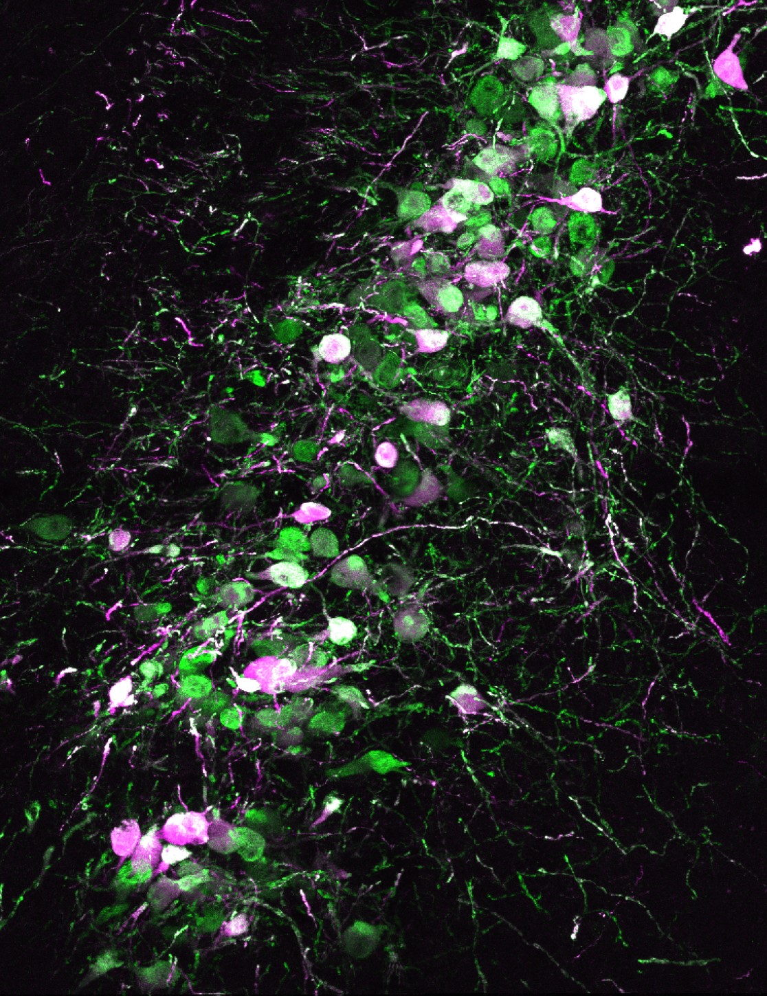
As described by Dr. Natale Sciolino, Will Fan, and Christopher Engborg: This image shows the anatomy of the adult mouse locus coeruleus (LC). The LC is a cluster of norepinephrine-synthesizing cells that help us attend to important sensory information. To monitor LC activity during tasting in behaving mice, a viral genetic strategy was used to express a calcium indicator (green) and a red fluorescent protein (magenta) in the LC and changes in fluorescence were recorded using fiber photometry.
In this image, a 40 µm-thick coronal section of a mouse LC was stained for the calcium sensor GCaMP8m (anti-GFP) and tdTomato reporter (anti-dsRed). The section was imaged using a 40x objective on a confocal microscope (A1R, Nikon). The cre-dependent GCaMP8m and tdTomato were both expressed in a subset of LC neurons in Dbhcre knock-in mice, with co-expression shown in white.
Using in vivo fiber photometry calcium recordings, the authors characterized LC’s unique activity patterns associated with licking behavior, shedding new light on its function.
Read the full article:
Locus Ceruleus Dynamics Are Suppressed during Licking and Enhanced Postlicking Independent of Taste Novelty
Will Fan, Christopher B. Engborg, and Natale R. Sciolino
FOLLOW US
 RSS Feed
POPULAR POSTS
TAGS
CATEGORIES


