Snapshots in Neuroscience: C. elegans PVD neuron dendrites
These images have been selected to showcase the art that neuroscience research can create.
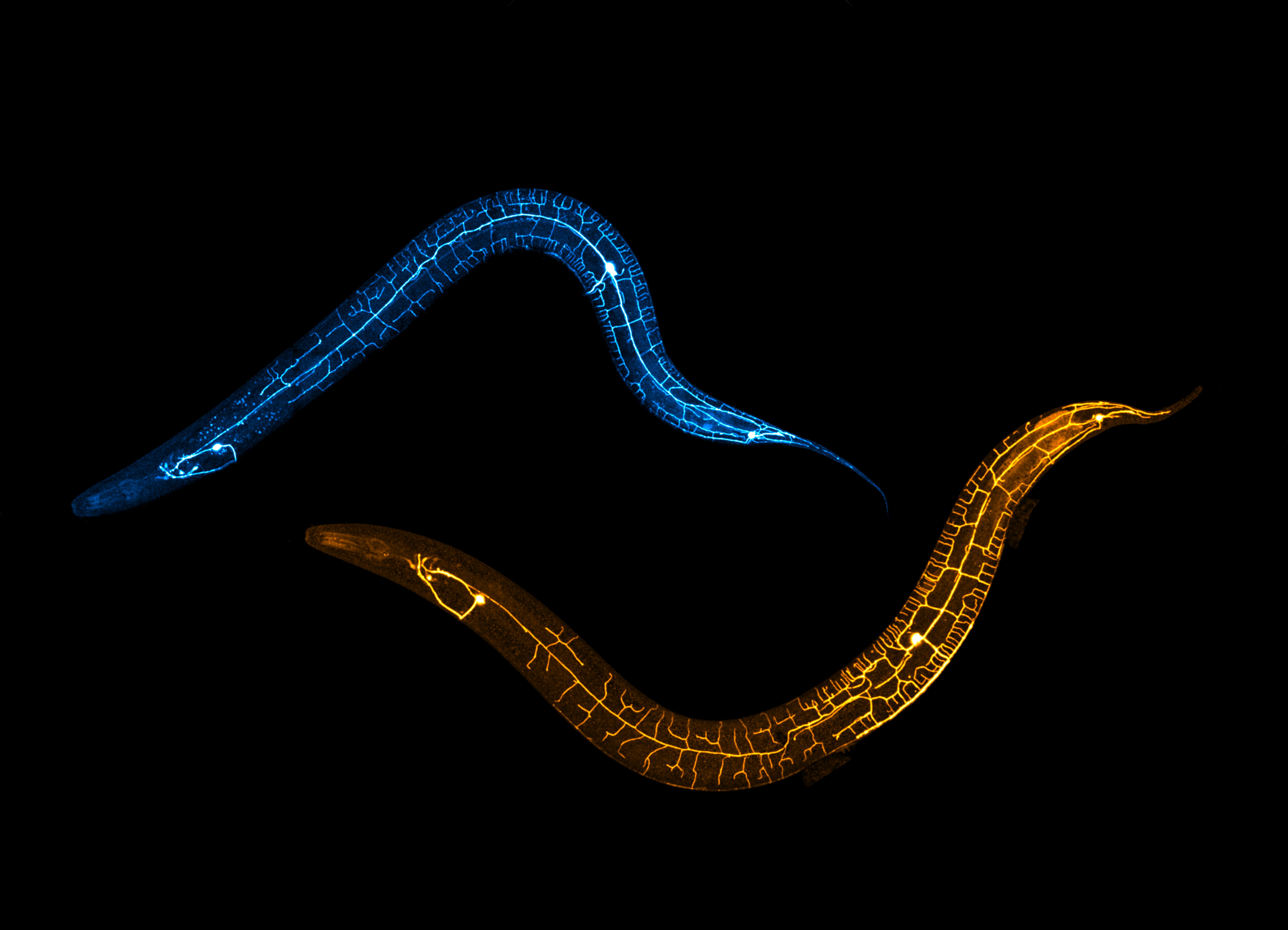
As described by Dr. Anindya Ghosh-Roy and colleagues: The PVD neuron of C. elegans controls multiple behaviors such as nociception and proprioception. The integrity of PVD dendrites governs proprioception in worms. This confocal image shows PVD neurons in C. elegans, with regenerating dendrites in golden hues and uninjured dendrites in cyan.
This image highlights the distinct regenerative pattern of PVD dendrites. Our study connects regenerative neurite growth to sensory behaviors, showing that axon regrowth impacts nociceptive touch, while dendrite regeneration affects posture.
The image was captured using confocal microscopy at 60X magnification with a fluorescent reporter expressed in PVD neurons to discern the neuronal structures. Z-stack images acquired at 60X magnification were tiled and stitched to align with the worm's body. Uninjured and regenerating PVD neurons were pseudocolored and combined into a single montage.
Read the full article:
Functional Recovery Associated with Dendrite Regeneration in PVD Neuron of Caenorhabditis elegans
Harjot Kaur Brar, Swagata Dey, Pallavi Singh, Devashish Pande, and Anindya Ghosh-Roy
FOLLOW US
 RSS Feed
POPULAR POSTS
TAGS
CATEGORIES


