Snapshots in Neuroscience: Mouse Cochlea
This image has been selected to showcase the art that neuroscience research can create.
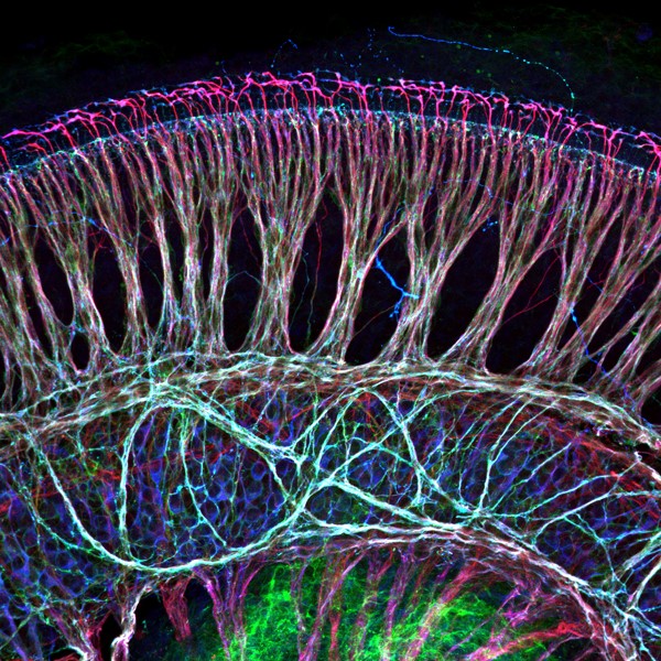
As described by Dr. Zhirong Wang and colleagues: This image shows an immunostained whole-mount preparation of a mouse cochlea. Cells localized at the bottom (blue) of the image are spiral ganglion neurons (primary auditory neurons) in the inner ear. Their peripheral axons bundle up (magenta) and extend towards sensory hair cells (not shown in this picture) to pick up sound information then relay it to the brain. There are also nerve fibers coming from the brain to modulate auditory input and they appear green/white in this image which covers the area of the neuronal cell body area.
Proper development of spiral ganglion neurons and precise connection between them and sensory hair cells are critical for hearing. We showed that a type of ATP-gated ion channels is required to maintain the size and complexity of spiral ganglion neuron terminal structures during early development.
Read the full article:
The Purinergic Receptor P2rx3 is Required for Spiral Ganglion Neuron Branch Refinement during Development
Zhirong Wang, Johnny S. Jung, Talya C. Inbar, Katherine M. Rangoussis, Christian Faaborg-Andersen and Thomas M. Coate
FOLLOW US
 RSS Feed
POPULAR POSTS
TAGS
CATEGORIES


