Snapshots in Neuroscience: Demyelinating Optic Nerve
This image has been selected to showcase the art that neuroscience research can create.
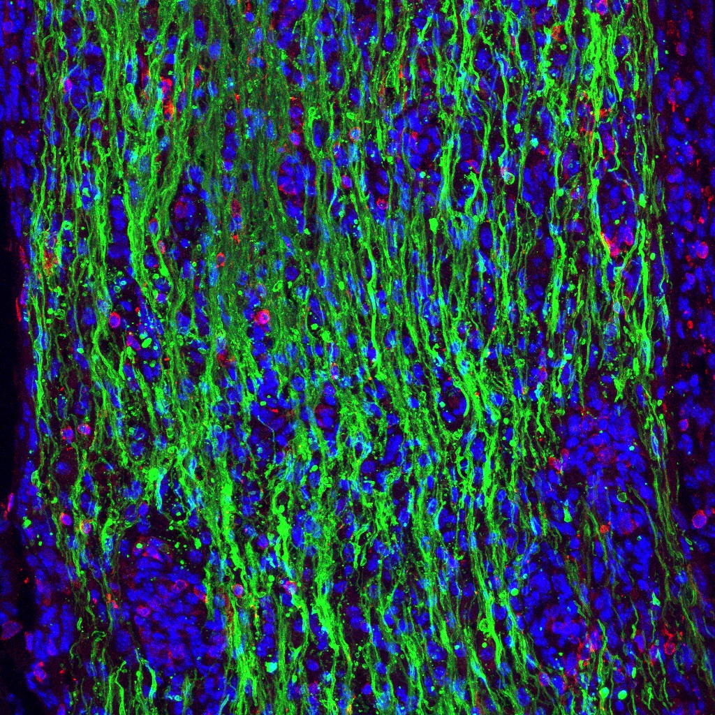
As described by Dr. Bhattacharya and Dr. Valdivia: This image shows the demyelinating optic nerve of an induced experimental autoimmune encephalomyelitis (EAE) mouse with impaired visual function. This is a 10 µm thickness section, stained for myelin basic protein (MBP) (green) which demonstrates the severity of demyelination that has occurred in this animal. Mature oligodendrocytes are stained in red by oligodendrocyte surface protein (OSP). DAPI staining of cell nuclei is shown in blue.
Optic neuritis, neuron-ectopic hyperdeimination, and demyelination are features of multiple sclerosis (MS) and in mouse models of demyelination, including EAE, lead to vision impairment. Our study identified a lyso-lipid (LPC 18:1) which can stem vision loss when injected sub-meningeal in the demyelinated optic nerve. LPC 18:1 promotes remyelination of the optic nerve by inducing oligodendrocyte maturation. Saturated lyso-lipids such as LPC 18:0 are demyelinating agents (used to develop the lysolecithin mouse model of demyelination). In contrast, our findings demonstrate that a single change in a double bond can have dramatic changes in biological function. Suggesting the potential therapeutic implications of exogenous delivery of LPC 18:1.
Our research has identified that a single bond change in a lyso-lipid results in a change in biology completely, that is from demyelinating (LPC 18:0) to a promoter of remyelination (LPC 18:1). This is perhaps the first example of a minor lipid structure (a single bond) change altering major biological function in neuronal or central nervous system.
Read the full article:
Lyso-Lipid-Induced Oligodendrocyte Maturation Underlies Restoration of Optic Nerve Function
Anddre Osmar Valdivia and Sanjoy K. Bhattacharya
FOLLOW US
 RSS Feed
POPULAR POSTS
TAGS
CATEGORIES


