Snapshots in Neuroscience: Mouse Brain Sagittal Section
This image has been selected to showcase the art that neuroscience research can create.
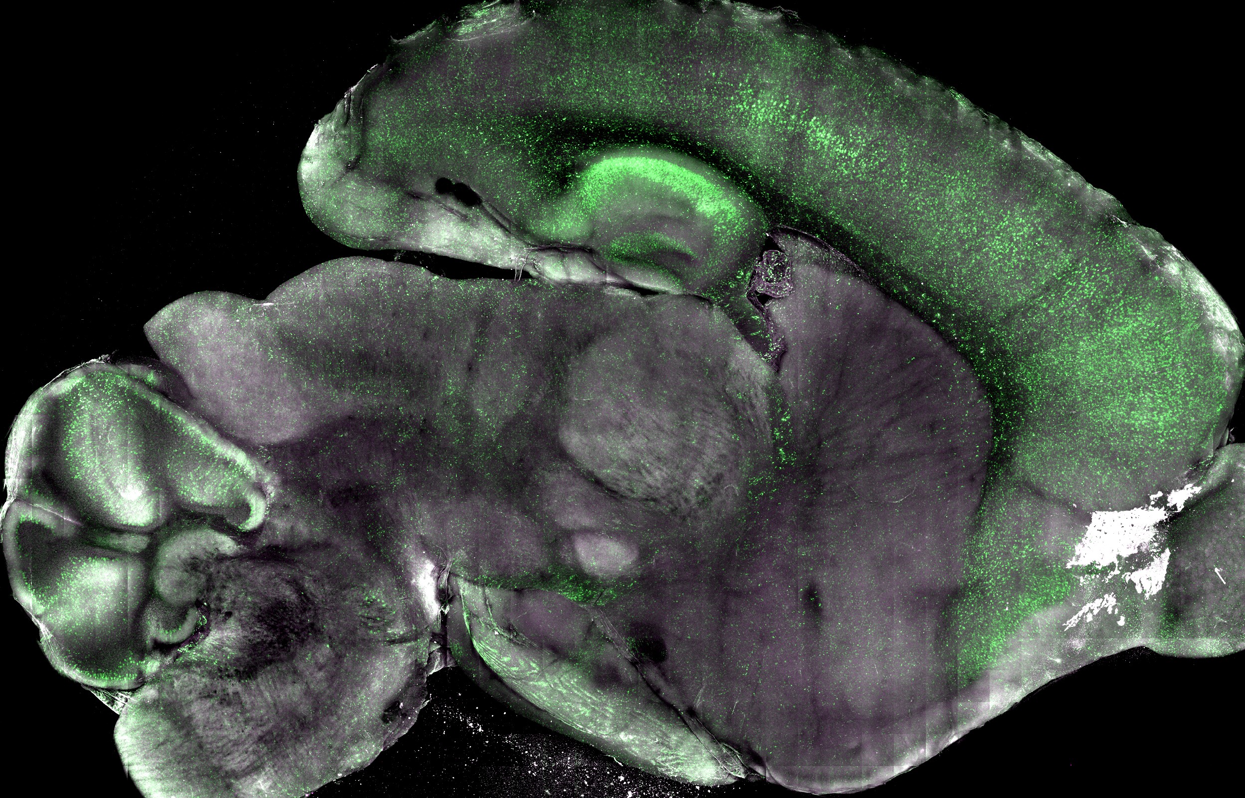
As described by Dr. Peter Nicholls and colleagues: The image shows a clarified, 1-mm-thick sagittal section of whole brain from a 22-day-old transgenic mouse expressing a fluorescent reporter for caspase-3/7 activity. In this sample, we were investigating the baseline level of reporter signal in a healthy, otherwise wild-type mouse.
You can note elevated reporter signal in the cortex, hippocampus, and cerebellar Purkinje cells; conversely, there is a relative lack of baseline signal in subcortical areas. We used multiphoton microscopy and a 20x/1.0 NA objective to perform a daylong scan at 3.3-micron resolution.
Green signifies reporter signal, while magenta represents autofluorescence; the two colors blend together to form the gray that predominates in the subcortical regions.
Read the full article:
Measuring Nonapoptotic Caspase Activity with a Transgenic Reporter in Mice
P. J. Nicholls, Thomas F. Pack, Nikhil M. Urs, Sunil Kumar, Yang Zhou, Gabriel Ichim, Joshua D. Ginzel, Gabor Turu, Evan Calabrese, Wendy L. Roberts, Ping Fan, Valeriy G. Ostapchenko, Monica S. Guzman Lenis, Flavio Beraldo, Jiri Hatina, Vania F. Prado, Marco A. M. Prado, Ivan Spasojevic, Joshua C. Snyder, Kafui Dzirasa, G. Allan Johnson, and Marc G. Caron
FOLLOW US
 RSS Feed
POPULAR POSTS
TAGS
CATEGORIES


