Snapshots in Neuroscience: Cerebral Neocortex
This image has been selected to showcase the art that neuroscience research can create.
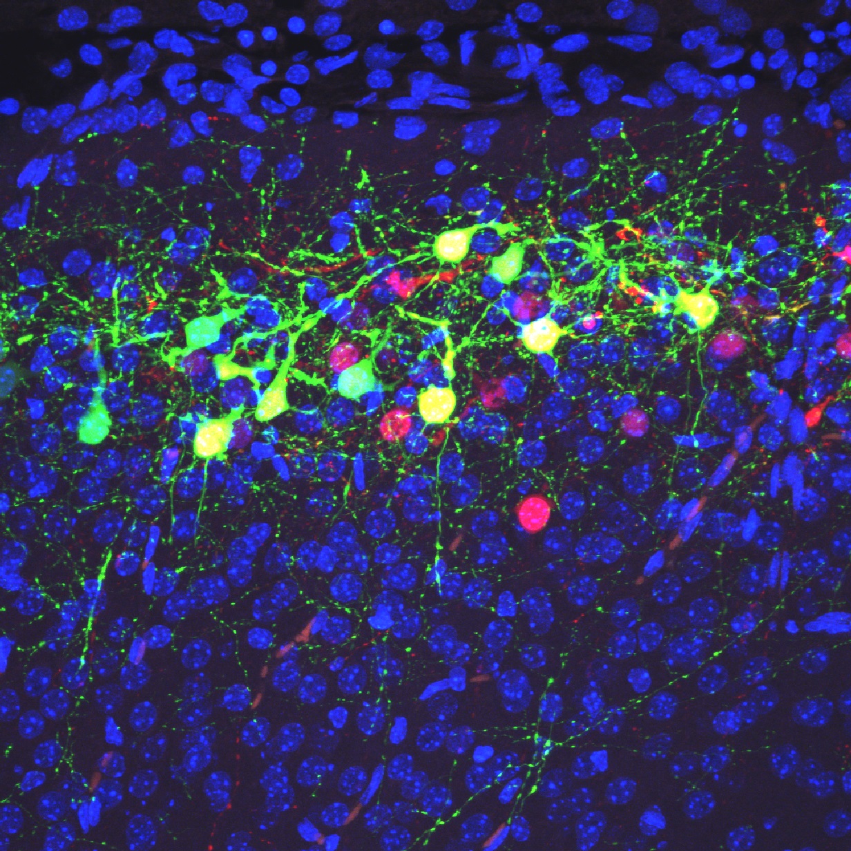
As described by Dr. Honda and colleagues: Green cells in this image are neurons distributed in the superficial region of the cerebral neocortex at postnatal day 9 in a heterozygous mutant mouse of Dab1, which plays an essential role in neocortex formation. Cells were labeled by electroporation of pCAGGS-RG and pDCX-Cre at embryonic day 16.5. pCAGGS-RG has a CAG promoter, a DsRed and a polyA signal flanked by two loxP sites, and an EGFP and a polyA signal in tandem, and normally expresses DsRed (magenta).
On the other hand, pDCX-Cre expresses Cre under the control of a Doublecortin (DCX) promoter. When pDCX-Cre and pCAGGS-RG are simultaneously introduced into a neuron, the neuron-specific DCX promoter would express Cre, which removes the DsRed and polyA cassette from pCAGGS-RG, resulting in EGFP (green) expression instead.
With a sparse labeling method using a combination of these plasmids, we found that in heterozygous mutants of Dab1, apical dendrites of superficial neurons are distributed more in neocortical layer 2 than in layer 1 compared to the wild-type mouse.
Read the full article:
Heterozygous Dab1 Null Mutation Disrupts Neocortical and Hippocampal Development
Takao Honda, Yuki Hirota, and Kazunori Nakajima
FOLLOW US
 RSS Feed
POPULAR POSTS
TAGS
CATEGORIES


