Snapshots in Neuroscience: Inhibitory Cortical Interneurons
This image has been selected to showcase the art that neuroscience research can create.
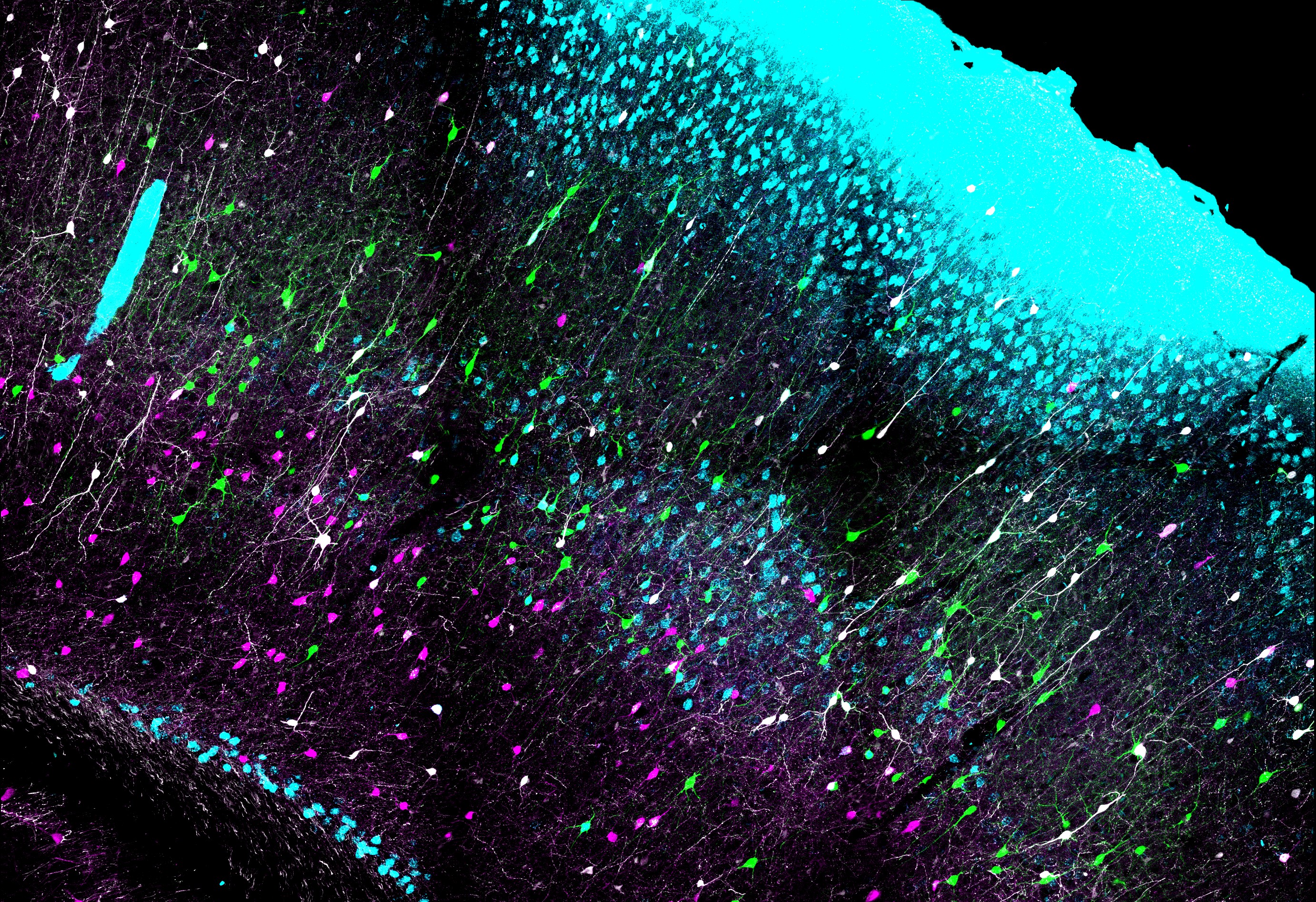
As described by Rachel E. Hostetler and colleagues: This image shows a coronal section of the somatosensory (barrel) cortex, from a triple-transgenic Sst-Flp;Pdyn-Cre;RC::FLTG mouse. In this mouse, Flp recombinase is expressed by all somatostatin-containing inhibitory interneurons, and Cre recombinase is expressed by cells expressing prodynorphin. When crossed with the dual color reporter (RC::FLTG), somatostatin interneurons that also express prodynorphin co-express a green fluorescent protein (GFP, green) while all other somatostatin interneurons (but no other neurons) express a red fluorescent protein (tdTomato,magenta). Prodynorphin-expressing somatostatin interneurons (green) were most highly concentrated in layers 4-5. To determine if this subset of interneurons sends axonal projections to layer 1 as do many somatostatin interneurons, we placed a deposit of Fast Blue dye on the pial surface, allowing 24 hour survival for the dye to be picked up by axon terminals and transported retrogradely to the cell bodies. Nearly half of the prodynorphin-expressing somatostatin interneurons, were retrogradely labeled by the dye (light blue), but very few were retrogradely labeled in layer 4. We also tested by immunostaining if prodynorphin-expressing somatostatin interneurons co-expressed several protein markers of cortical interneurons such as calretinin (white).
We repeated these experiments on triple-transgenic crosses with three other Cre driver lines, characterized these different subsets of somatostatin interneurons electrophysiologically, and compared them with previously identified somatostatin subtypes. Our data reveal three non-overlapping subtypes of somatostatin-containing interneurons with different genetic labels, electrophysiological characteristics, laminar distributions of cell bodies and axons terminals, and protein marker expression. Together, these three subtypes account for >50% of somatostatin interneurons in layer 5 and for >40% of the total somatostatin interneuron population.
Read the full article:
Genetically Defined Subtypes of Somatostatin-Containing Cortical Interneurons
Rachel E. Hostetler, Hang Hu, and Ariel Agmon
FOLLOW US
 RSS Feed
POPULAR POSTS
TAGS
CATEGORIES


