Snapshots in Neuroscience: BDNF expression
This image has been selected to showcase the art that neuroscience research can create.
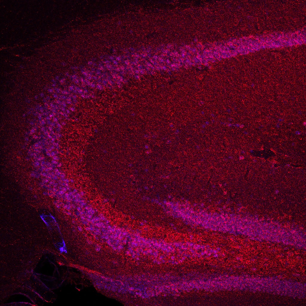
As described by Dr. Linden and Ms. Janowitz: This image depicts a slice from the brain of a mouse cut in the sagittal plane. The immunofluorescence shows expression of brain-derived neurotrophic factor (BDNF) in the CA3 region and parts of the dentate gyrus and hilus of the hippocampus following chronic treatment with the serotonin selective reuptake inhibitor (SSRI) sertraline. The slice was immunolabeled for BDNF (red) and neuronal nuclear antigen (NeuN) (blue) to identify neuronal cell bodies.
Treating mice daily for 6 weeks with 10 mg/kg of sertraline increased levels of BDNF in the CA3 region and hilus of the hippocampus compared to saline-treated controls. In our study, we investigated the effect of SSRIs on the regrowth of serotonin axons in the adult forebrain following a chemical injury. Although there was no effect of sertraline in our model, the increase in BDNF, shown here, served as a positive control for our method of administration.
This image is a maximum intensity projection of a 40 μm thick tissue slice captured using confocal microscopy at 120x magnification.
Read the full article:
Chronic treatment with serotonin selective reuptake inhibitors does not affect regrowth of serotonin axons following amphetamine injury in the mouse forebrain
Haley Janowitz and David Linden
FOLLOW US
 RSS Feed
POPULAR POSTS
TAGS
CATEGORIES


