Snapshots in Neuroscience: Hippocampal astrocytes and radial glial stem cells
These images have been selected to showcase the art that neuroscience research can create.
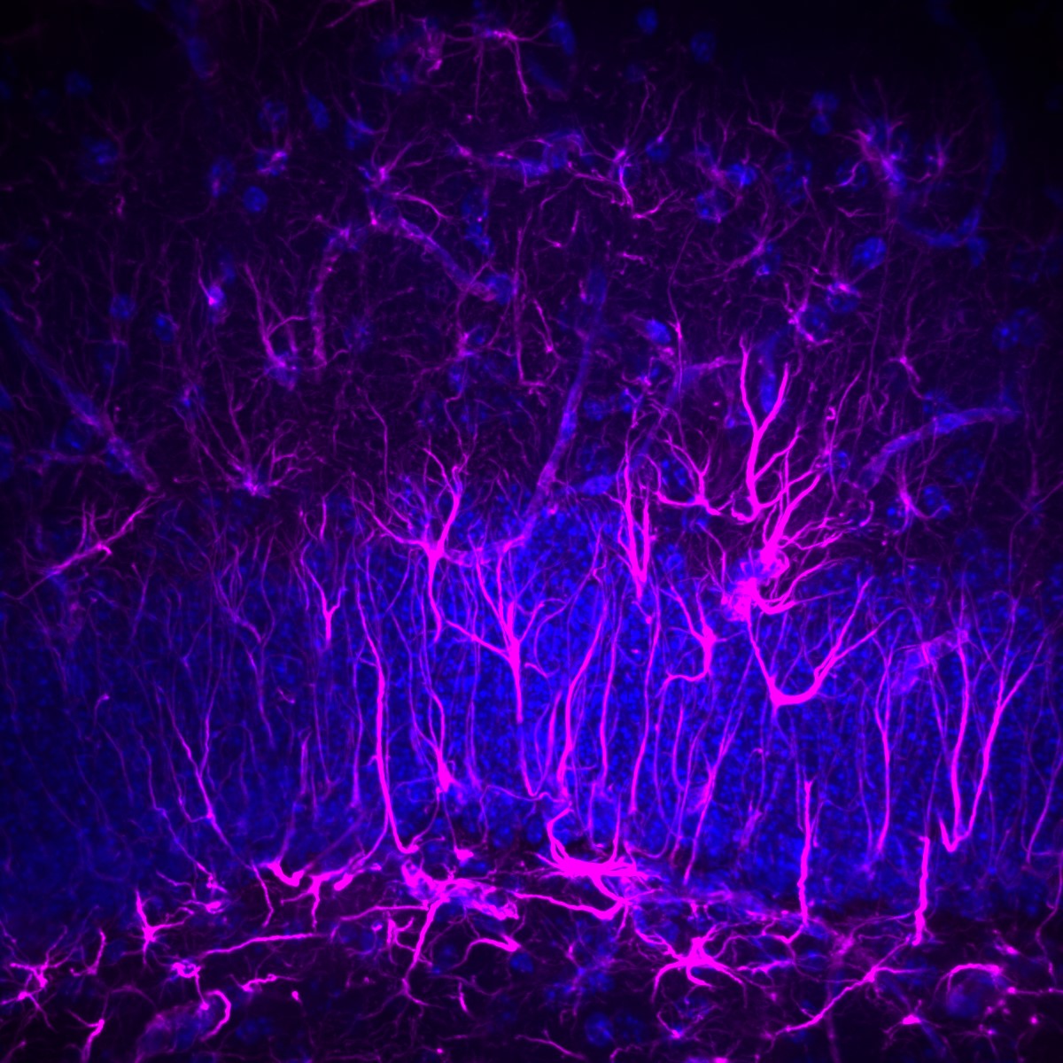
As described by Dr. Schnell and colleagues: This image was obtained from a section of the mouse hippocampal dentate granule cell layer (highlighted here with DAPI staining in blue) after immunohistochemical staining for Glial Fibrillary Acidic Protein (GFAP; magenta), which is expressed by astrocytes and radial glial stem cells. It is a maximum projection of a 3-dimensional image Z-stack, obtained on a Spinning Disk Confocal microscope through a 40x 1.4NA oil objective. GFAP-stained astrocytes have a typical swept-spider morphology, while radial glial cells extend radially through the granule cell layer.
Images like this one derive from an in-depth characterization of hippocampal dentate gyrus histopathology in CACNA2D2 knockout mice, which are epileptic and prone to handling-induced seizures. Although most structural and cellular correlates of temporal lobe epilepsy were absent in the dentate gyri of these mice, they exhibited dramatic changes in granule cell activation in response to both handling and low-dose chemoconvulsants, as measured by c-fos and ∆FosB expression. Together, these findings indicated clear functional changes in the hippocampal circuit without (perhaps yet) identified structural correlates.
Read the full article:
Altered Hippocampal Activation in Seizure-Prone CACNA2D2 Knock-out Mice
Alyssa B. Danis, Ashlynn A. Gallagher, Ashley N. Anderson, Arielle Isakharov, Kathleen A. Beeson, and Eric Schnell
FOLLOW US
 RSS Feed
POPULAR POSTS
TAGS
CATEGORIES


