
Snapshots in Neuroscience: Mouse Traumatic Brain Injury
These images have been selected to showcase the art that neuroscience research can create.
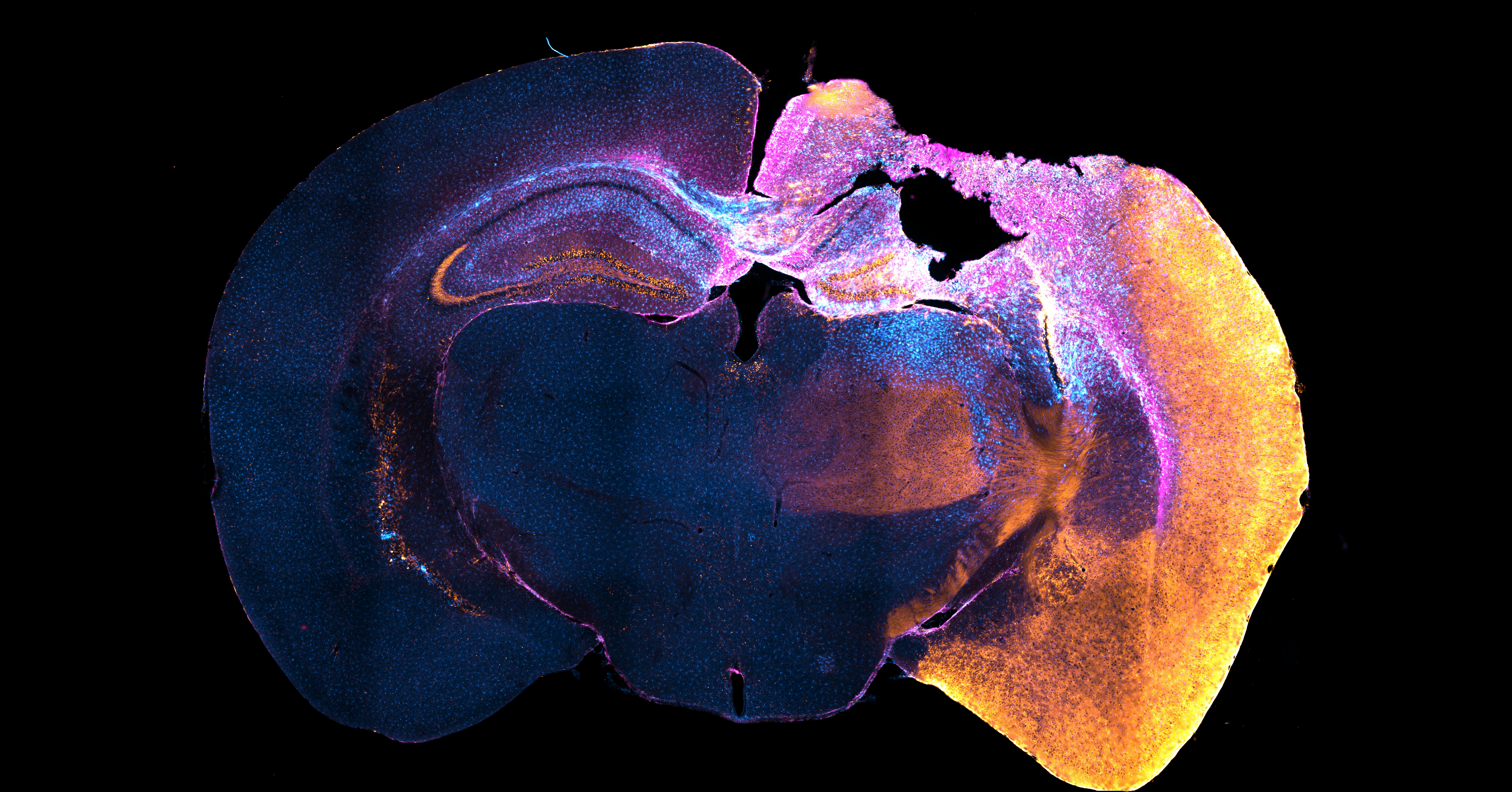
As described by the authors: “This image is a coronal brain section showing all the neurons that were activated after a traumatic brain injury. The mouse is a Targeted Recombination in Active Populations (TRAP2) Cre driver crossed to an Ai14 reporter line. This genetic mouse line uses a tamoxifen-inducible system tied to the Fos gene to permanently label neurons that become active shortly after a contusive brain injury. We stained this section for tdTomato to reveal activated neurons (orange, Ai14) as well as GFAP (magenta, astrocytes) and IBA1 (blue, microglia) to mark the injury site.
“In our study, we used TRAP2-based whole-brain mapping of Ai14 expression in the early postinjury period. This revealed that cortex-wide neuron activation occurs shortly after a focal brain injury, including a near complete engagement of the ipsilateral hemisphere. We also noted substantial Ai14 expression in amygdala, internal capsule, cerebral peduncle, and lateral thalamic nuclei known to interact with somatosensory neocortex, including reticular thalamus and ventrobasal complex. These brain areas likely become engaged during the primary response to injury.
“Immunostaining was performed on 50 μm coronal brain sections. The image was acquired using a Leica DM6 microscope at 5× magnification.”
Read the full article:
Cortex-Wide Neuron Activation after Traumatic Brain Injury in Mice
Alexa Tierno, Aidan Baraban, and Robert F. Hunt
FOLLOW US
 RSS Feed
POPULAR POSTS
TAGS
CATEGORIES