
Snapshots in Neuroscience: Dentate Hilius Neurons
These images have been selected to showcase the art that neuroscience research can create.
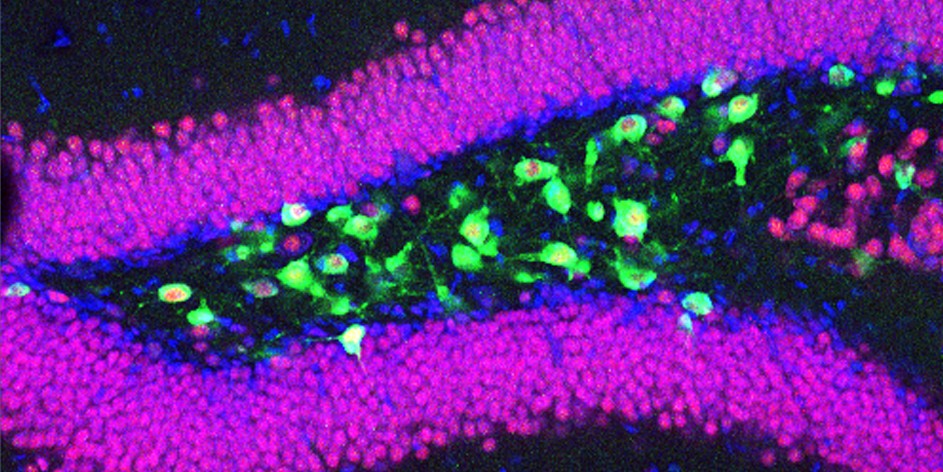
As described by the authors: “This confocal image shows the dentate hilus of the hippocampus from a Drd2-Cre:Eef1a1LSL.EGFP-L10a/+ mouse. In this transgenic mouse line, Cre recombinase drives expression of the EGFP-L10a ribosomal fusion protein specifically in Drd2-expressing neurons. Cell nuclei are labeled with DRAQ5 (blue), mature neurons are identified by NeuN immunostaining (red), and EGFP-L10a fluorescence labels Drd2-expressing hilar neurons (green), a population composed primarily of mossy cells with a smaller subset of interneurons.
“In our study, we used Translating Ribosome Affinity Purification and RNA sequencing to characterize translatomic features of Drd2-expressing hilar neurons along the dorsoventral axis of the hippocampus. Although these neurons appear anatomically similar, we found that dorsal and ventral populations are molecularly distinct. These findings suggest that dorsoventral molecular heterogeneity among hilar neurons provides new insights into functional specialization.
“Immunostaining was performed on 40 µm cryosections using antiNeuN antibody (rabbit polyclonal, 1:1,000, #ABN78, Millipore). Image was acquired as collapsed 4 µm Z-stacks using a Nikon confocal microscope with a 20× objective.”
Read the full article:
Translational Profiling of Drd2-Expressing Populations Reveals Molecular Heterogeneity of Dentate Gyrus Mossy Cells along the Dorsoventral Axis
Minseok Jeong, Jin-Hyeok Jang, Seo-Jin Oh, Ji-Woong Choi, and Yong-Seok Oh
FOLLOW US
 RSS Feed
POPULAR POSTS
TAGS
CATEGORIES