Snapshots in Neuroscience
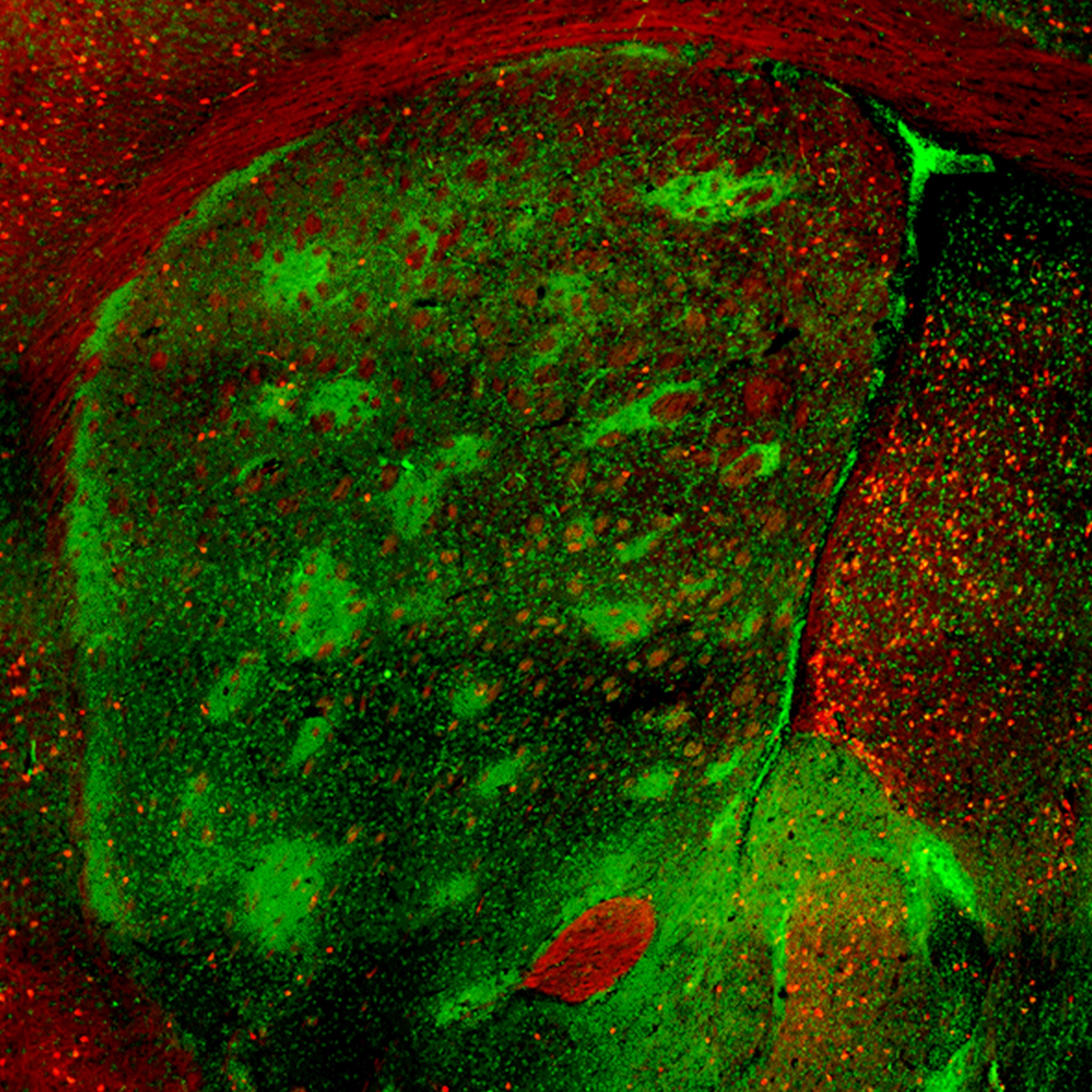
Neurons in the male mouse striatum that are activated by social vocalizations during courtship.
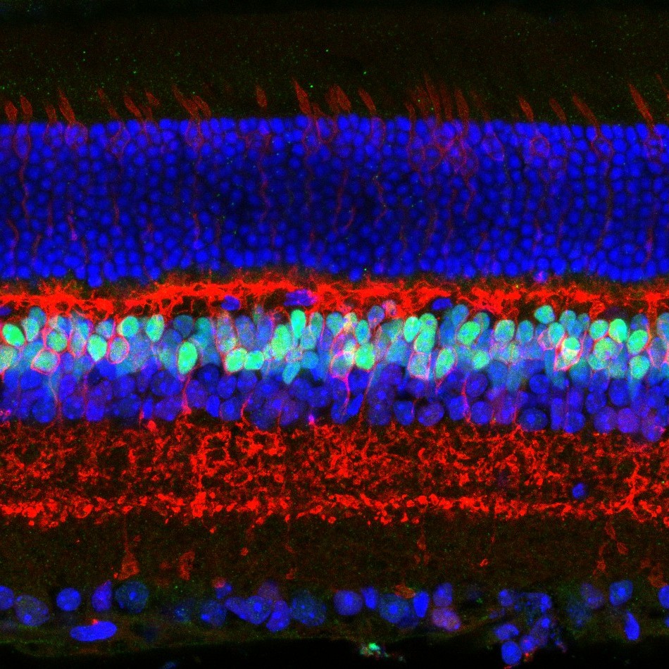
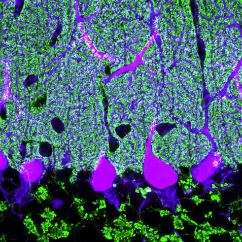
Bipolar cells and cone photoreceptor cells in the inner and outer nuclear layers of the adult mouse retina.
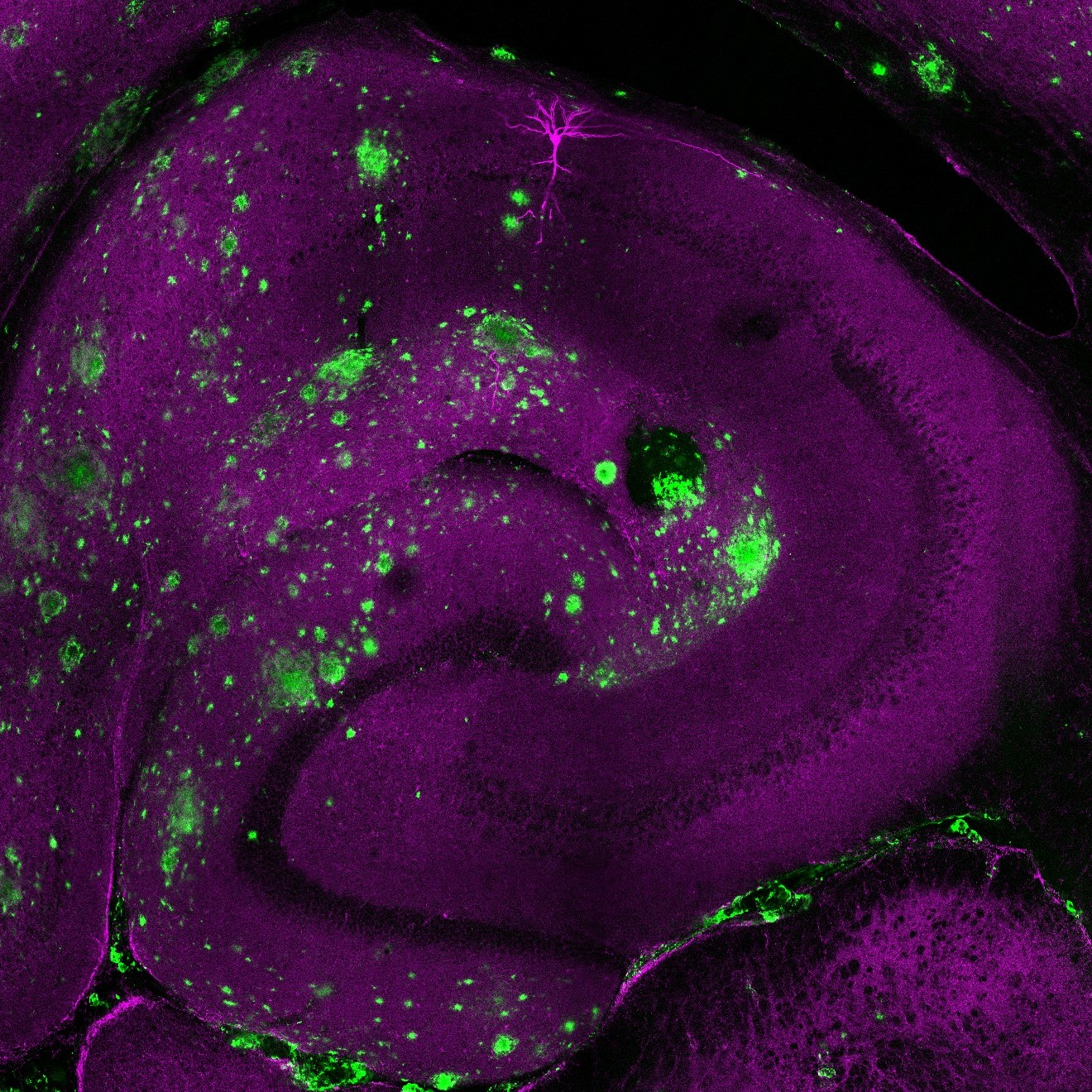
Neurotoxic plaques and a CA1 pyramidal neuron in the hippocampus of an Alzheimer’s disease mouse model.
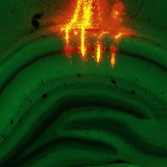
Vivid vertical tracks targeting the Cornu Ammonis 1 region of the dorsal hippocampus of a transgenic rat.
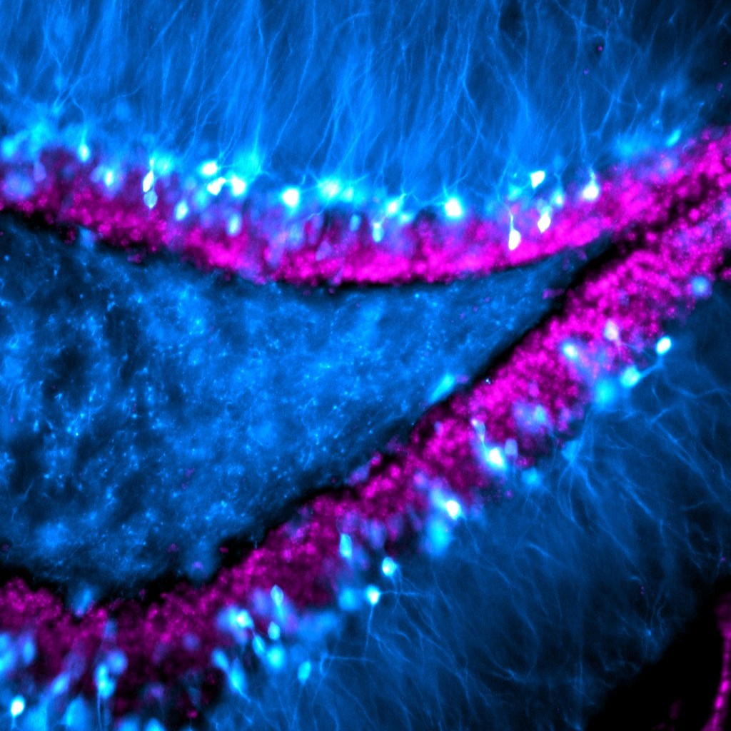
Granule cells in the dentate gyrus of a mouse, with visible extensions of their dendrites reaching out into the molecular layer.
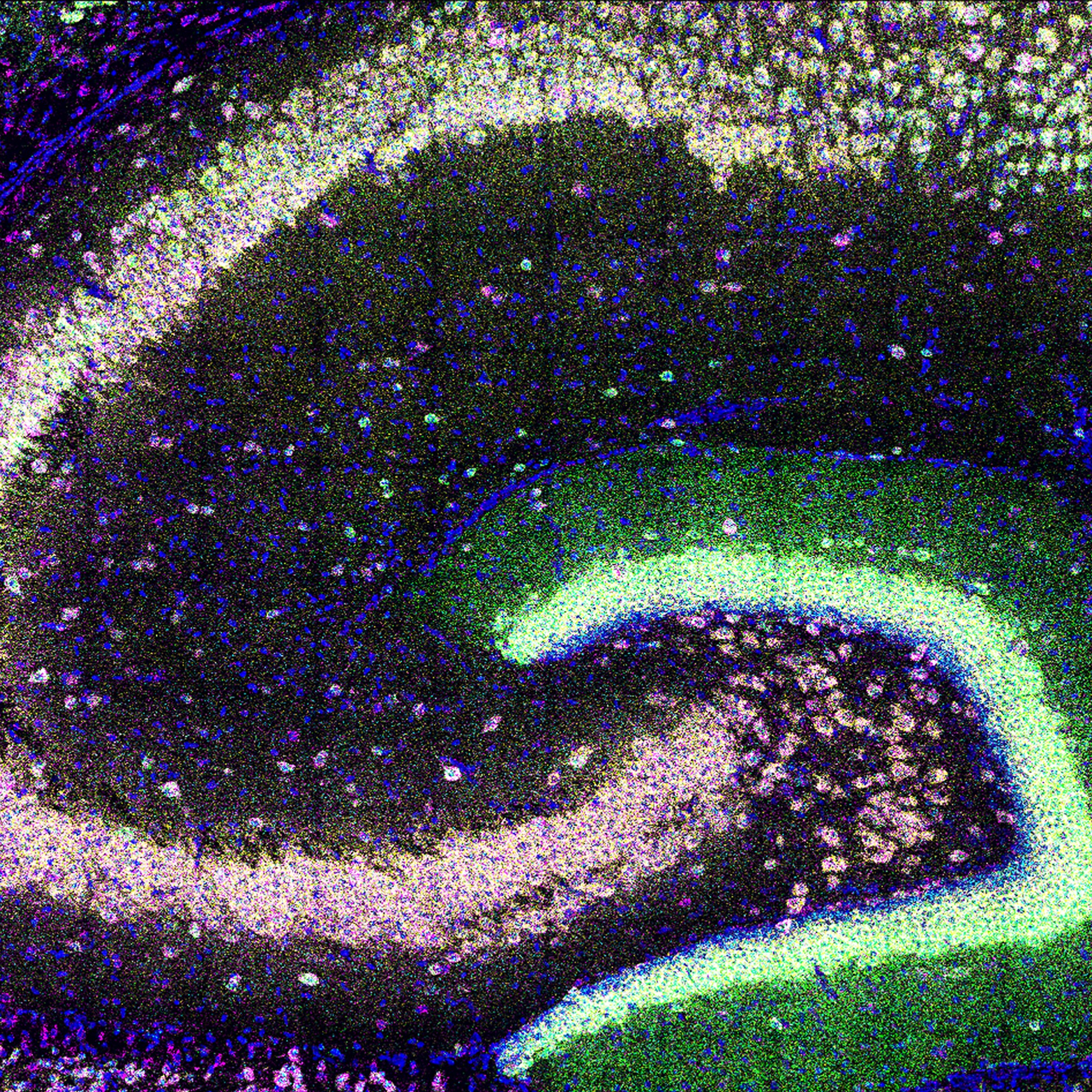
Expression of four neuropil-localized mRNA in hippocampal subregions of a young mouse.
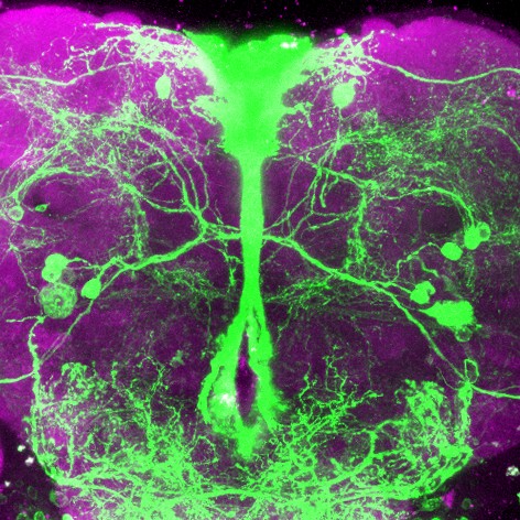
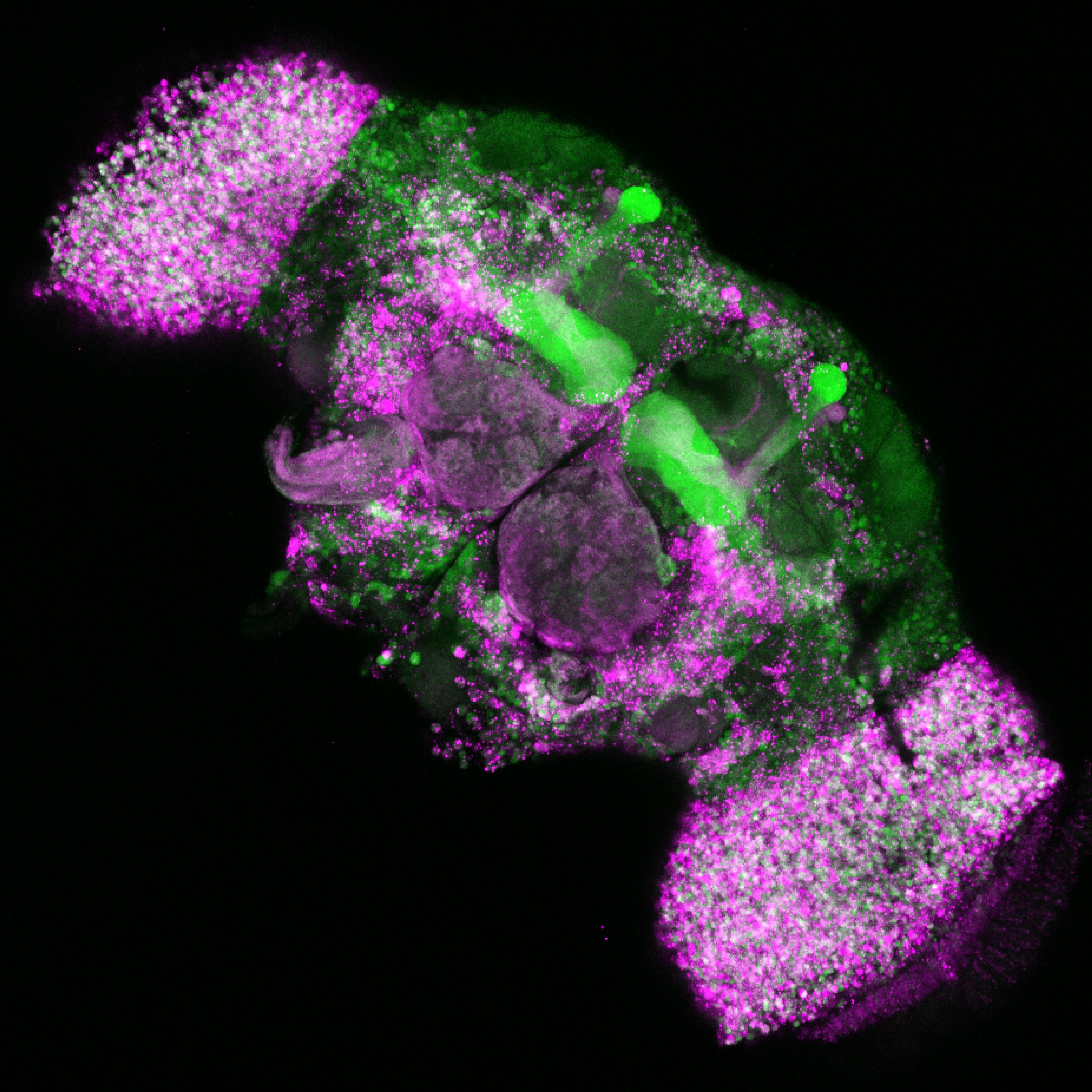
Mapping neuropeptide F receptor expressing-neurons and their role in thirsty water seeking in the adult male fruit fly.
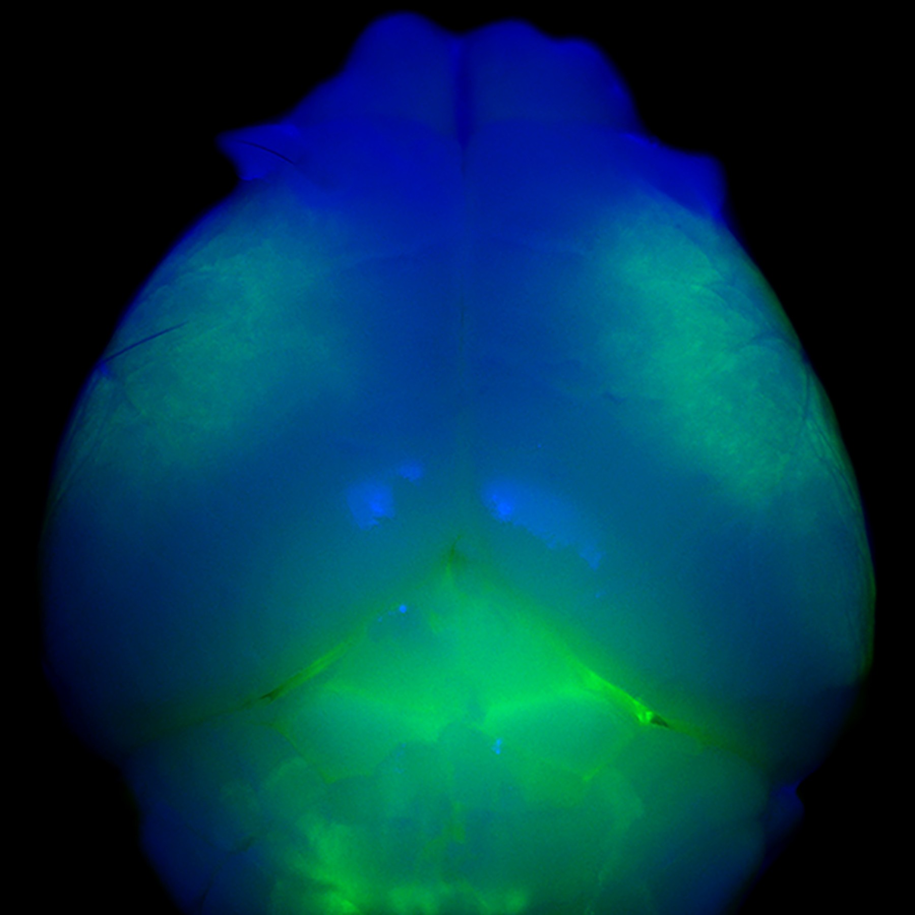
Whole mount view of a young mouse brain with all cortical layer 5 neurons expressing dystonia-related gene Klhl14, highlighted in green.
Neurons in the adult fruit fly brain have different biases in the splicing of a calcium channel that enables neurotransmission.
Mouse cerebellar section with a single layer of Purkinje cells extending their elaborate dendritic branches upwards into the dense molecular layer.
FOLLOW US
 RSS Feed
TAGS
CATEGORIES


