Disorders of the Nervous System
Looking back at ten years of eNeuro papers, this post features two papers published in 2019.
New evidence for sleep disruption as an early symptom of Alzheimer’s disease progression that accelerates cognitive decline in a sex-specific manner.
New genetic mouse model sheds light on the relationship between genetic predisposition and environmental triggers for OCD-like behaviors and paves the way for novel treatment strategies.
Looking back at ten years of eNeuro papers, this post features two papers published in 2015.
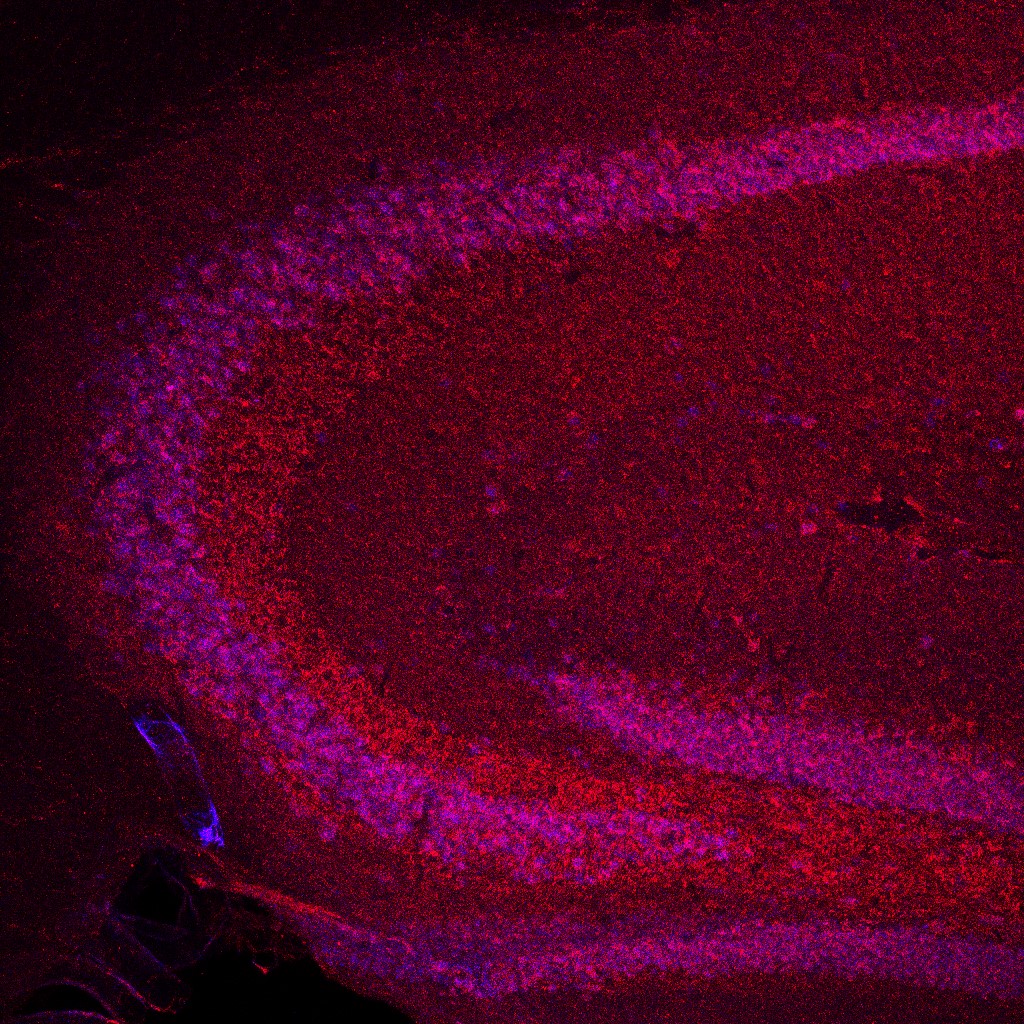
A maximum intensity projection image of a sagittal mouse brain slice captured using confocal microscopy.
Karl Herrup and Christophe Bernard philosophically discuss fallacy traps in neuroscience, in an episode of the webinar series SfN Journals: In Conversation. Here is a teaser for the episode available to watch on-demand.
Dr. Paul Baudin, currently completing his medical degree at the Sorbonne Paris Nord university, tells us how his Ph.D. broadened his conceptualization of neurological diseases and treatments, and emphasizes the importance of publishing negative data.
The authors explain how splicing variation (3R vs. 4R) and cytoplasmic tethering (by 14-3-3) blunt the pathogenicity of phosphorylated tau during normal brain development.
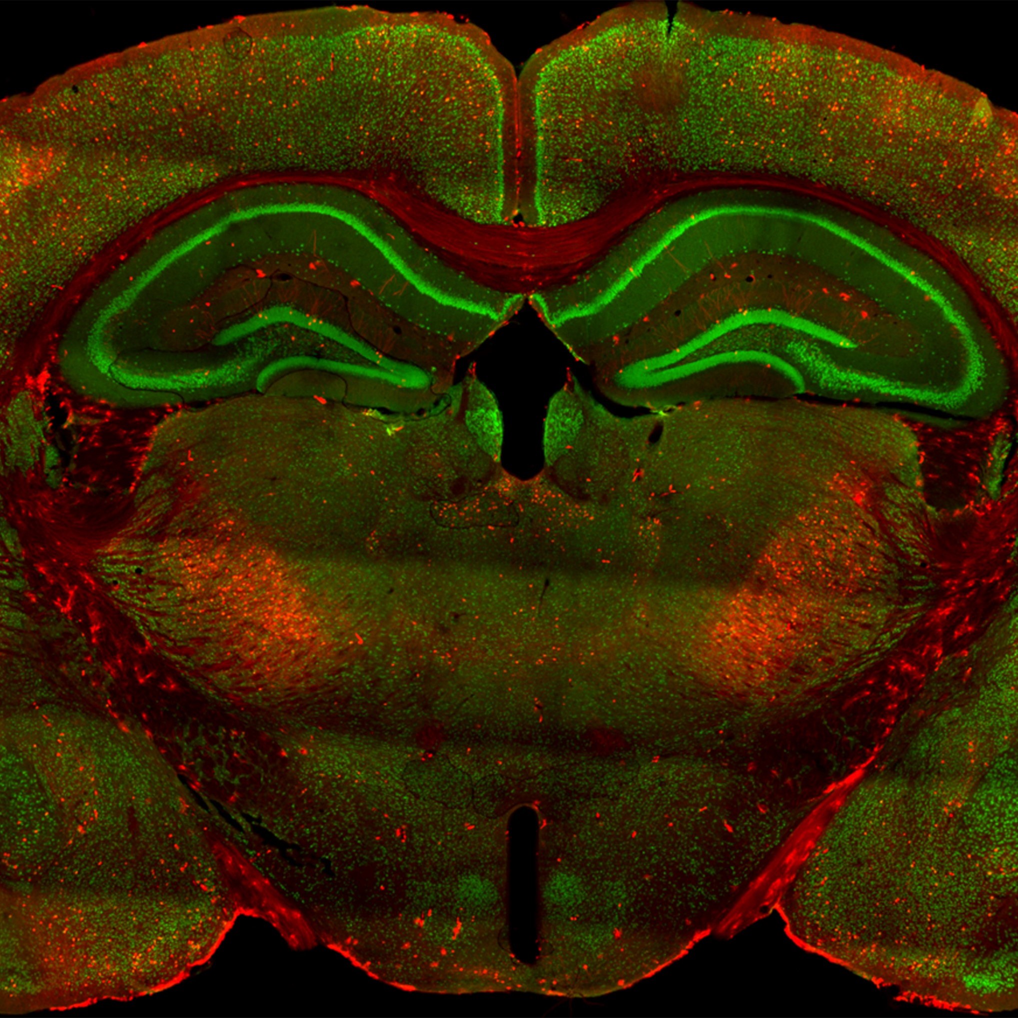
This image shows coronal slices at the level of the hippocampus in TRAP2 mice labeling neuronal ensembles during a rewarded T-maze spatial memory task.
This paper presents an elegant study examining neuronal activation in multiple brain regions involved in learning and consolidation of an alternation task.
FOLLOW US
TAGS
CATEGORIES


 RSS Feed


