Development
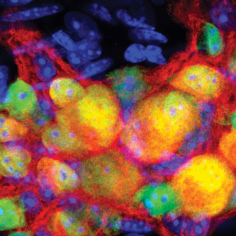
Immunostaining of cell bodies and nerve fibers of neurons in an adult mouse small intestinal myenteric ganglion.
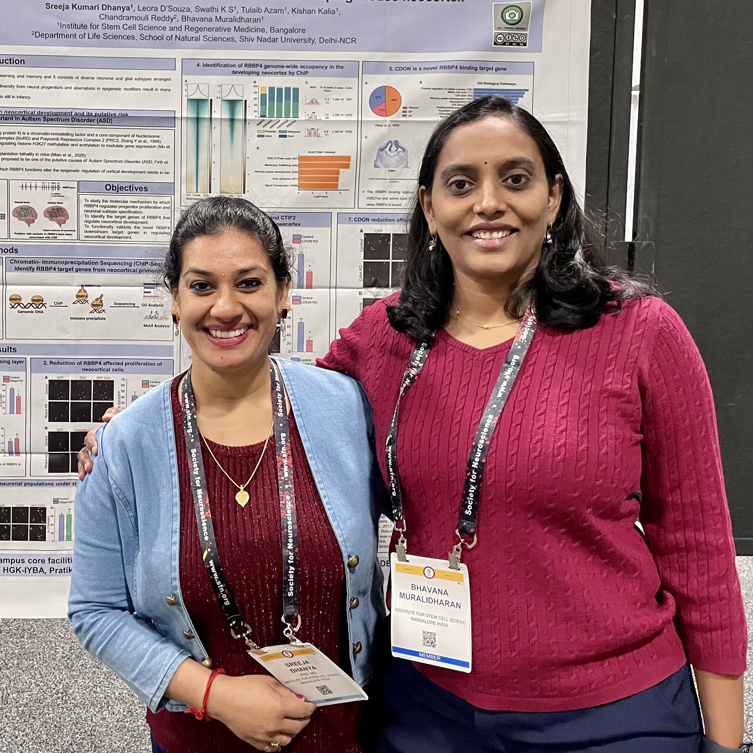
Drs. Sreeja Kumari Dhanya and Bhavana Muralidharan emphasize the value of supportive mentorship and a flexible mindset as they discuss their study unveiling a new role for a chromatin modifier in neurogenesis.
Looking back at ten years of eNeuro papers, this post features two papers published in 2023.
Looking back at ten years of eNeuro papers, this post features two papers published in 2022.
Looking back at ten years of eNeuro papers, this post features two papers published in 2019.
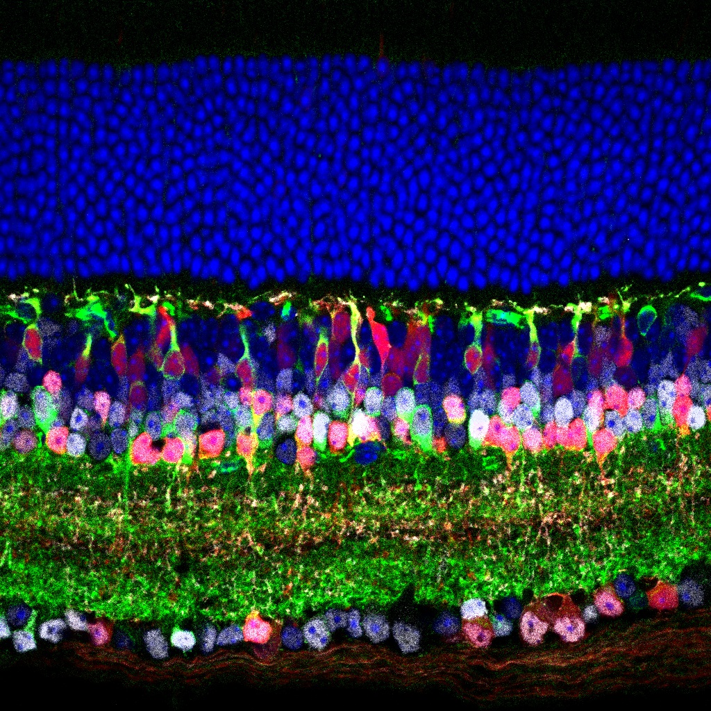
This image shows the cellular layers of an adult mouse retina, stained for markers of amacrine cells and type 3b bipolar cells.
In this study, authors find an important new locus of compensatory synaptic strength changes in the early developing chick autonomic nervous system.
This paper investigates spatial and temporal patterns of key enzymes involved in purine synthesis.

Dr. Matthew Colonnese gives behind-the-scenes details about his eNeuro paper and cogitates on the implications of his surprising findings, in an episode of the webinar series SfN Journals: In Conversation. Here we provide a teaser for the episode available to watch on-demand.
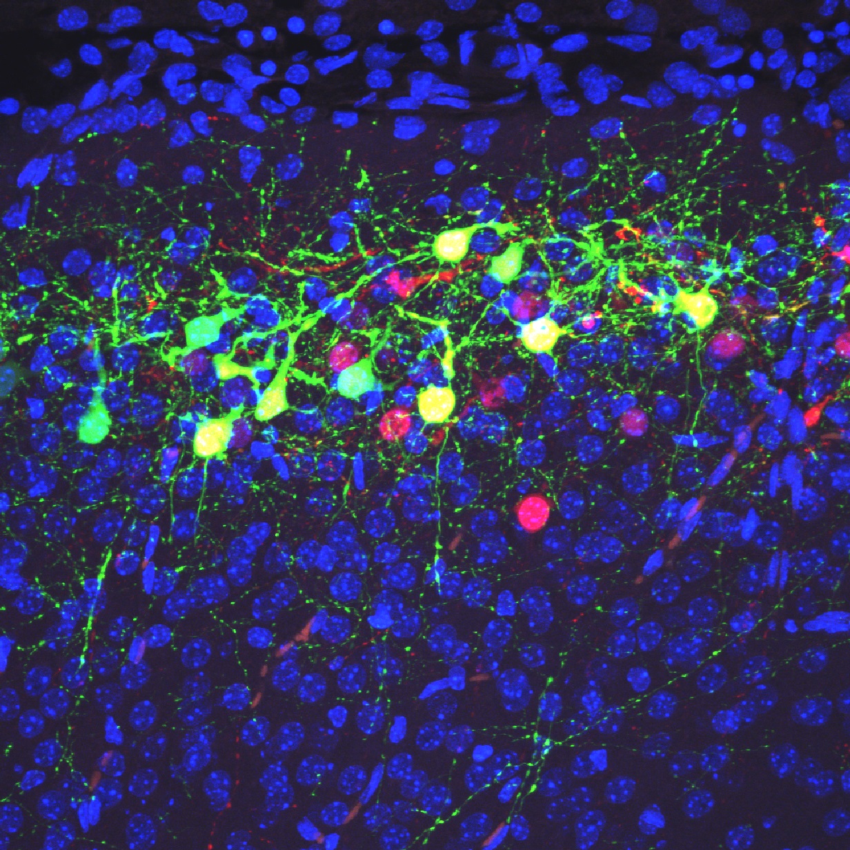
Green cells in this image are neurons distributed in the superficial region of the cerebral neocortex at postnatal day 9 in a heterozygous mutant mouse of Dab1.
FOLLOW US
 RSS Feed
TAGS
CATEGORIES


