Featured Finding

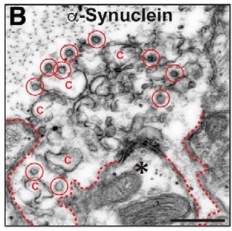
Image of electron micrographs of untreated, control lamprey synapses stimulated at 20 Hz for 5 min.
By Rosalind S.E. Carney, DPhil
July 9, 2020

Authors report a viable chaperone-based strategy for reversing the synaptic vesicle trafficking defects associated with excess α-synuclein, which may be of value for improving synaptic function in Parkinson’s disease and other synucleinopathies.

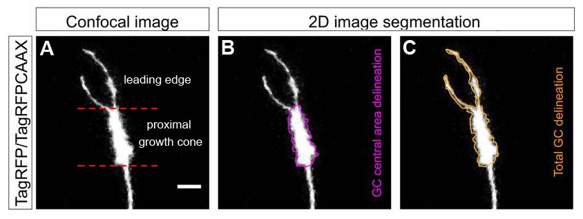
Authors describe the in vivo behavior of mitochondria at the growth cone of elongating retinal axons in zebrafish.
By Rosalind S.E. Carney, DPhil
February 6, 2020

Authors describe the in vivo behavior of mitochondria at the growth cone of elongating retinal axons in zebrafish.

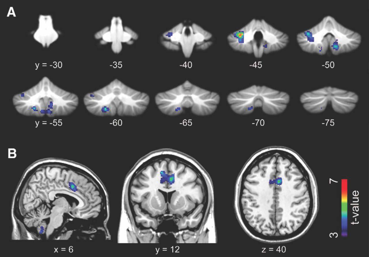
Authors show that real-time mutual interaction during eye contact is mediated by the cerebellum and limbic mirror system.
By Rosalind S.E. Carney, DPhil
October 10, 2019

Authors show that real-time mutual interaction during eye contact is mediated by the cerebellum and limbic mirror system.

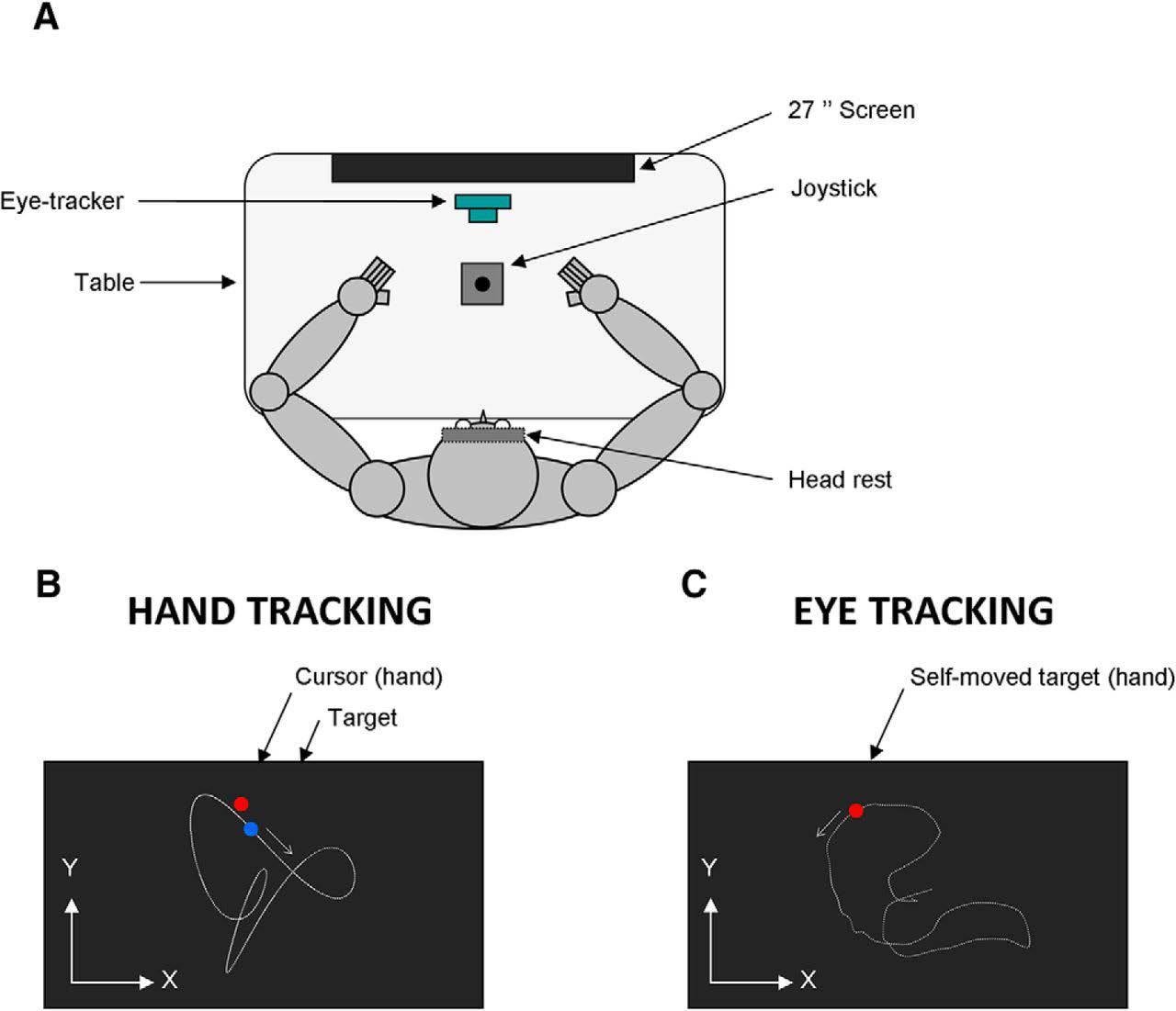
Authors show that although handedness impacts the accuracy of hand movement control, it has virtually no influence on the ability to predict the visual consequences of hand movements.
By Rosalind S.E. Carney, DPhil
August 8, 2019

Authors show that although handedness impacts the accuracy of hand movement control, it has virtually no influence on the ability to predict the visual consequences of hand movements.

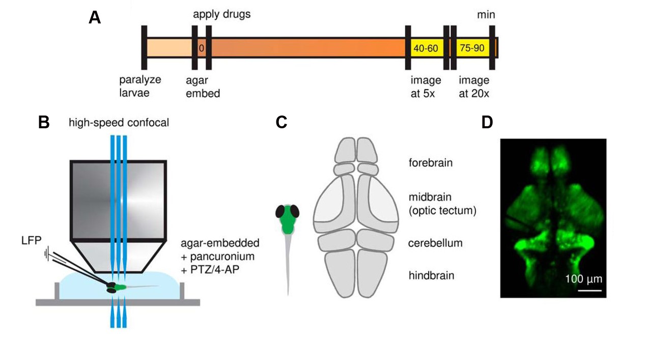
Figure 1. High-speed confocal imaging and LFP recording in the forebrain of GCaMP-expressing zebrafish larvae. (A) Timeline of confocal imaging and LFP recording relative to immobilization of larvae in agar and exposure to convulsant drugs. High-speed confocal recordings were obtained ∼40 minutes after drug exposure, with 5× and 20× objectives used for whole-brain and single neuron imaging, respectively. (B) Simultaneous high-speed confocal imaging and LFP recording in agar-embedded larvae. (C) Schematic illustration depicting sub-regions of the larval zebrafish brain. (D) Representative 5× image of Tg(neurod1:GCaMP6f) zebrafish at 5–6 days postfertilization. (Adapted from Figure 1 in Liu & Baraban et al., 2019.)
By Rosalind S.E. Carney, DPhil
May 9, 2019

Authors used well-established seizure protocols and fast confocal imaging of genetically encoded calcium indicator-expressing zebrafish to investigate epileptic network properties at brain-wide and single-cell levels.

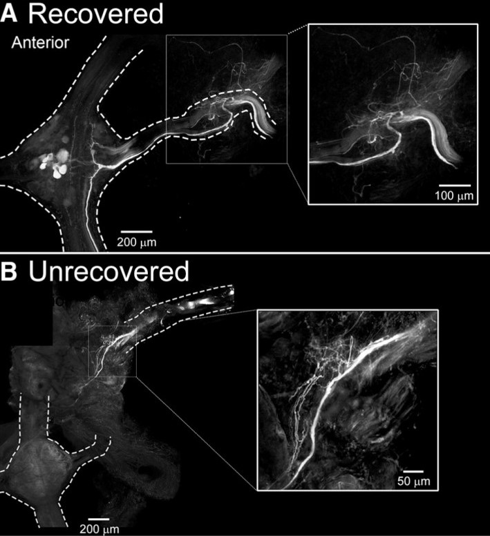
Figure 1. Leeches that recover crawling behavior have established regrowth of afferent input to the lead ganglion. (A) In leeches that recover crawling behavior after injury, Neurobiotin labels fibers in the M3 nerve, which project into the M3 ganglion. (B) In leeches that do not recover crawling behavior after injury, Neurobiotin-labeled fibers are not observed in the M3 ganglion. (Figure 8 in Puhl et al. 2018.)
By Rosalind S.E. Carney, DPhil
December 13, 2018

Authors demonstrate that in the absence of descending cephalic input, locomotor recovery in the leech is achieved by a switch to dependence on afferent information from peripheral nerves in the body wall.

By Rosalind S.E. Carney, DPhil
October 31, 2018

Authors demonstrate that capsaicin, a TRPV1 agonist, activates a pro-axon growth program, suggesting an approach for enhancing axon regeneration in selective populations of neurons.

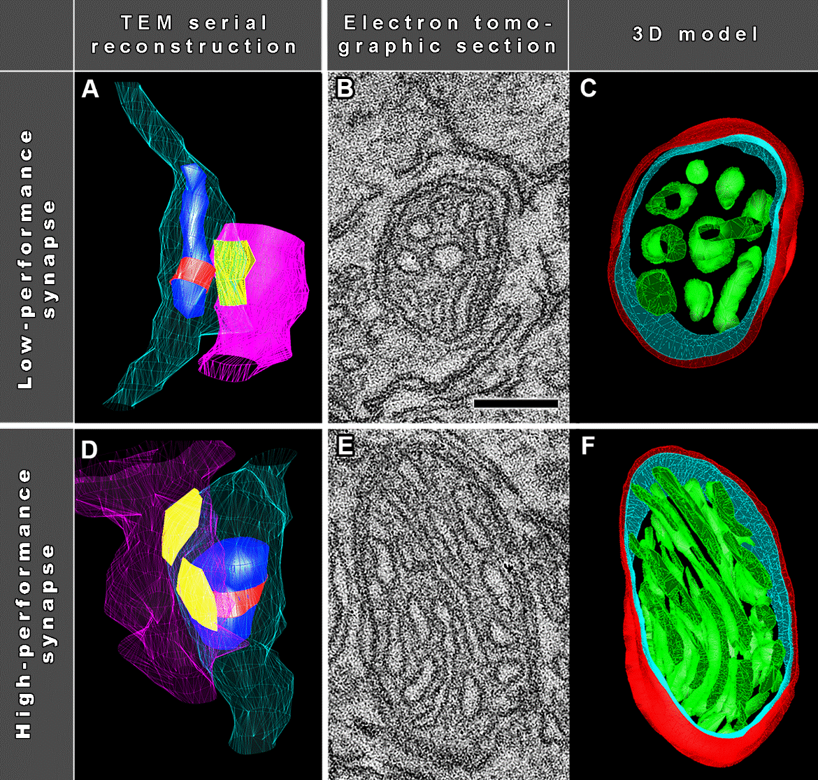
Mitochondrial Ultrastructure is Optimized for Synaptic Performance (Adapted from Figure 3 in Cserép et al., 2018)
By Rosalind S.E. Carney, DPhil
September 21, 2018

Authors find an evolutionarily conserved, cell-type independent coupling of mitochondrial ultrastructure to synaptic performance.