Sensory and Motor Systems
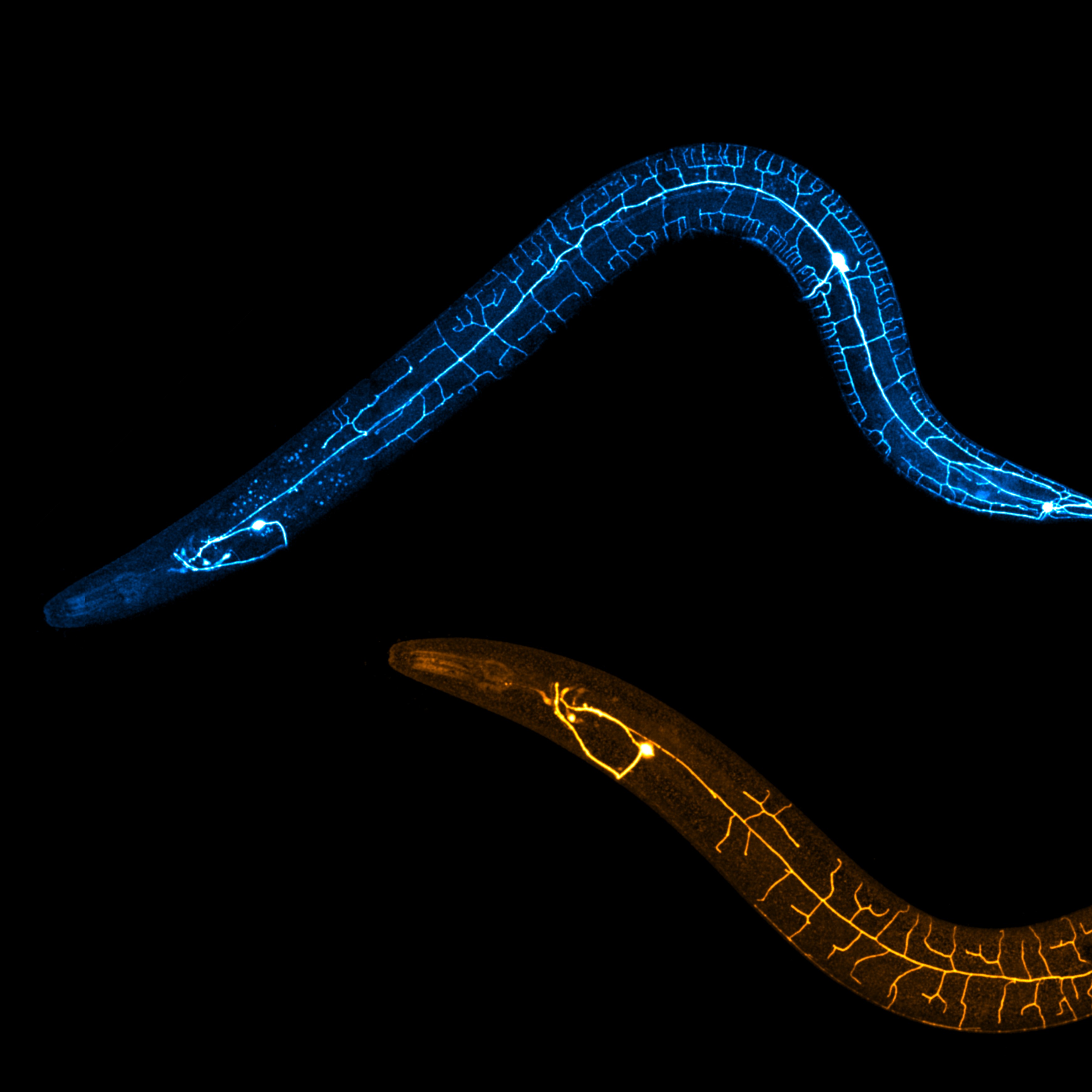
Shown are regenerating dendrites and uninjured dendrites of PVD neurons in C. elegans.
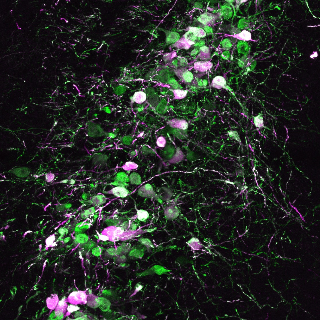
To assess neural activity during mouse tasting behavior, a calcium indicator (green) and a red fluorescent protein (magenta) were expressed in the locus coeruleus.
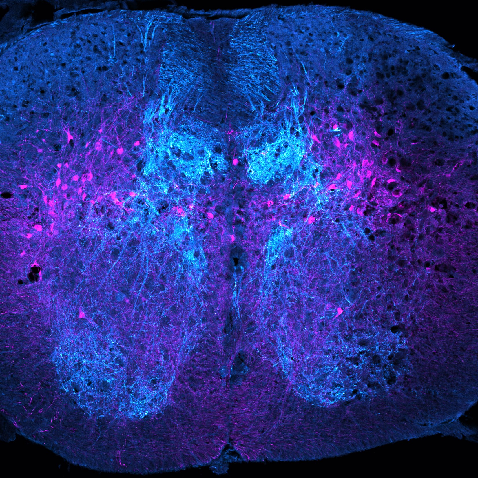
Pictured are proprioceptive sensory afferents and Atoh1-lineage neurons in the lower thoracic mouse spinal cord.
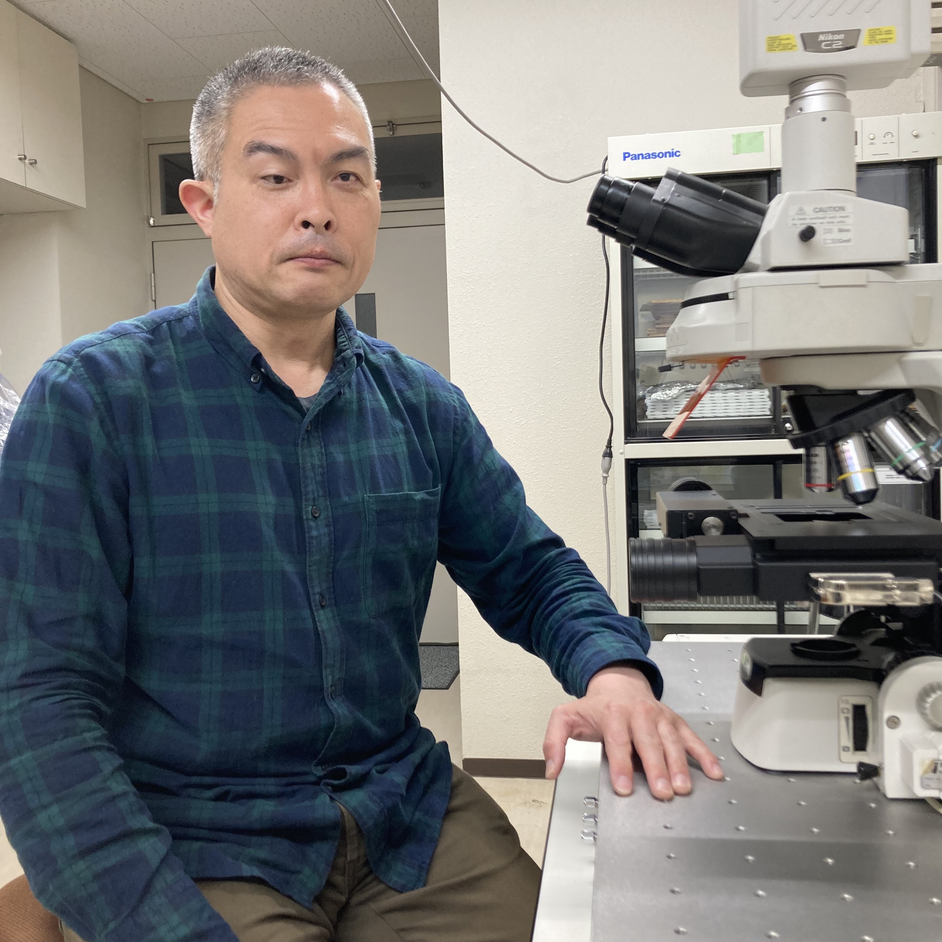
Dr. Naoki Shigematsu talks about how his research interests led him to Dr. Takaichi Fukuda’s lab where he investigated the under-explored anterolateral barrel subfield and discusses the findings of his paper.
Co-first authors Drs. Krishnakanth Kondabolu and Natalie Doig discuss how they overcame barriers from COVID lockdowns to collaborate, bridging together multiple techniques to map out and describe a precise connection between the subthalamic nucleus and the striatum.
Dr. Mahima Sharma, currently advancing her research career at the Buck Institute, emphasizes how interdisciplinary collaboration is critical for extending benchwork science translatability as we discuss her recent first author eNeuro publication.
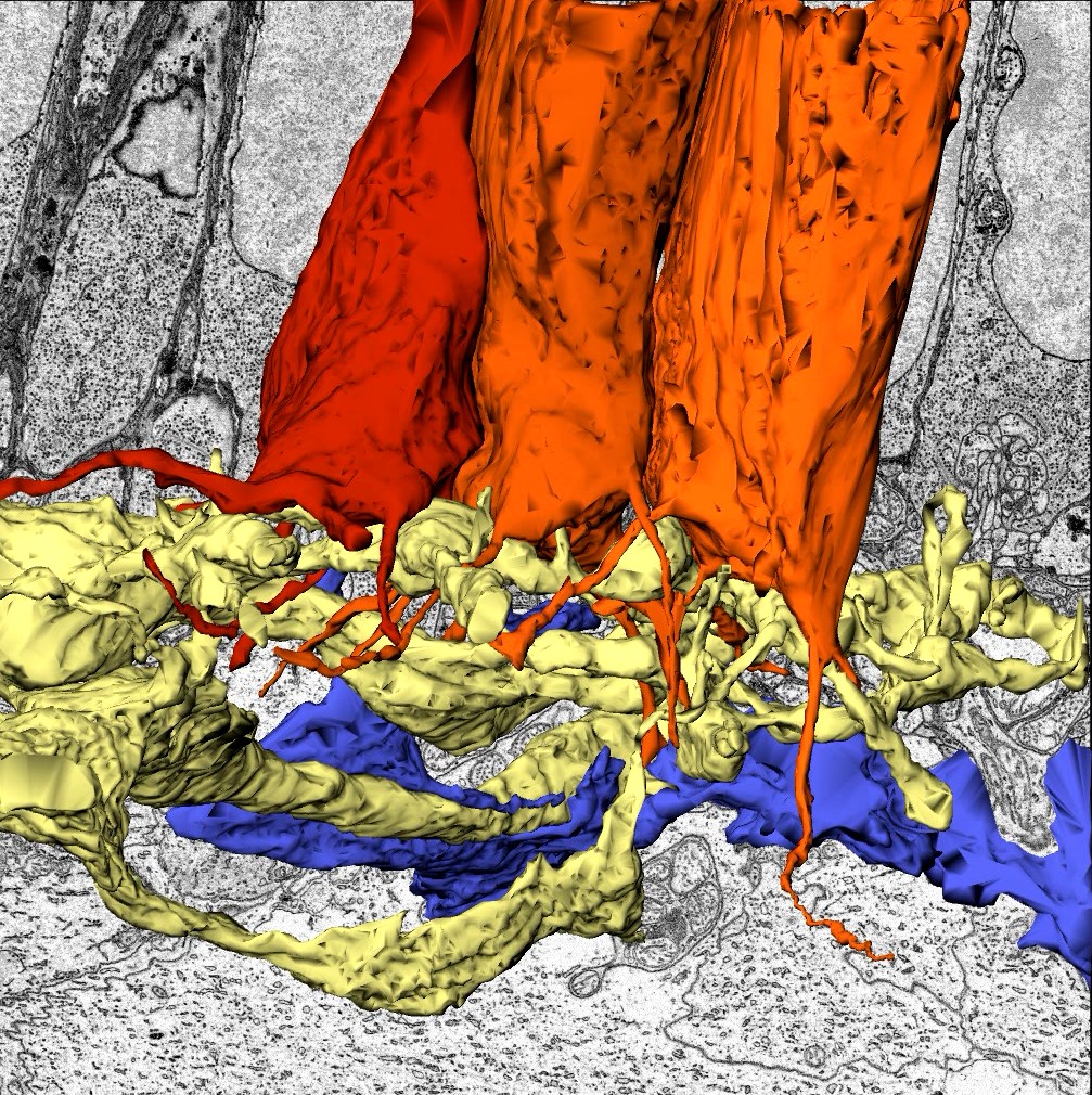
This image shows 3D reconstructions of Little skate photoreceptor terminals (orange) and postsynaptic partners (yellow and blue) obtained from serial block-face electron microscopy data.
Dr. Hugo Calligaro, currently in the lab of Dr. Satchidananda Panda at Salk Institute for Biological Studies, tells us about his interest in circadian rhythms and discusses the unexpected findings from his recent first author publication.
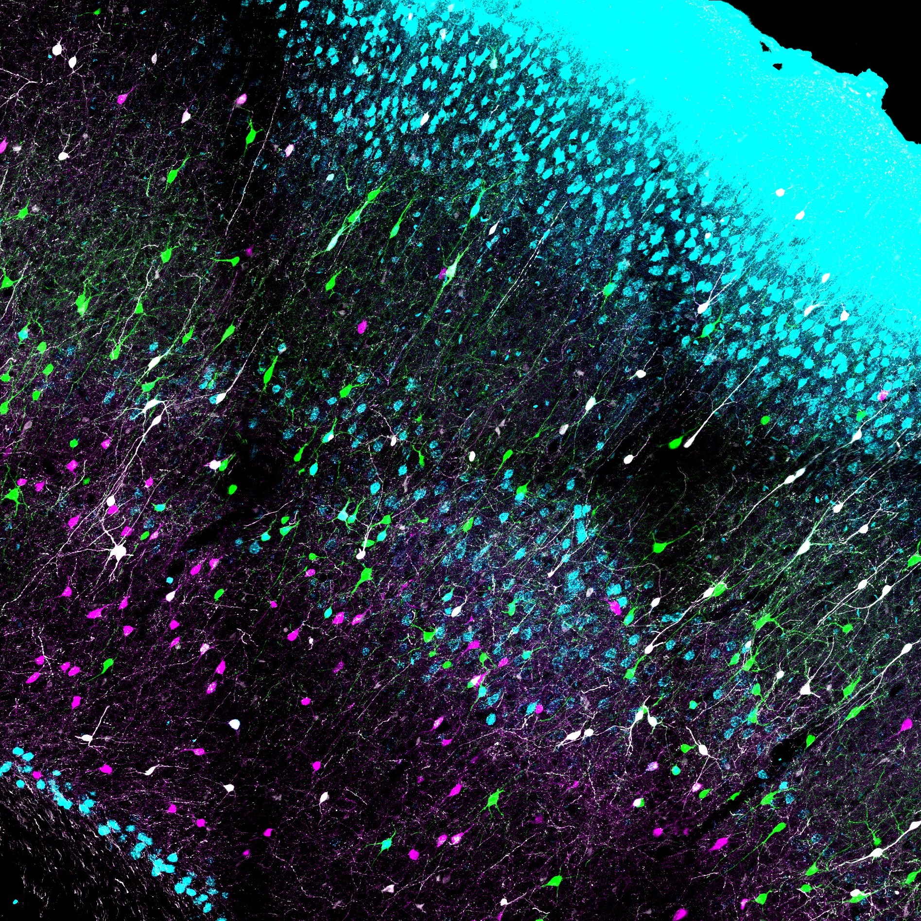
Confocal image of a brain section containing the somatosensory (barrel) cortex from a transgenic mouse.
The authors show that voltage-gated sodium channels in vomeronasal sensory neurons undergo slow inactivation and this can contribute to the spike adaption caused by repeated stimuli.
FOLLOW US
TAGS
CATEGORIES





 RSS Feed


