Snapshots in Neuroscience
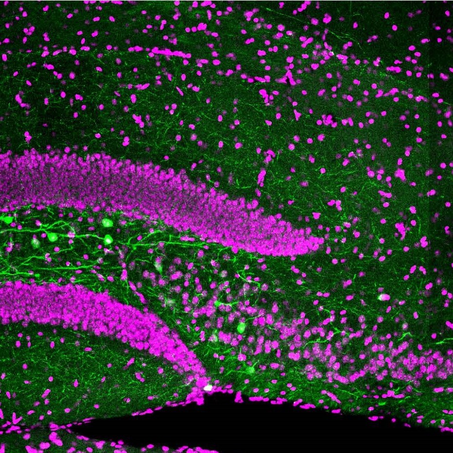
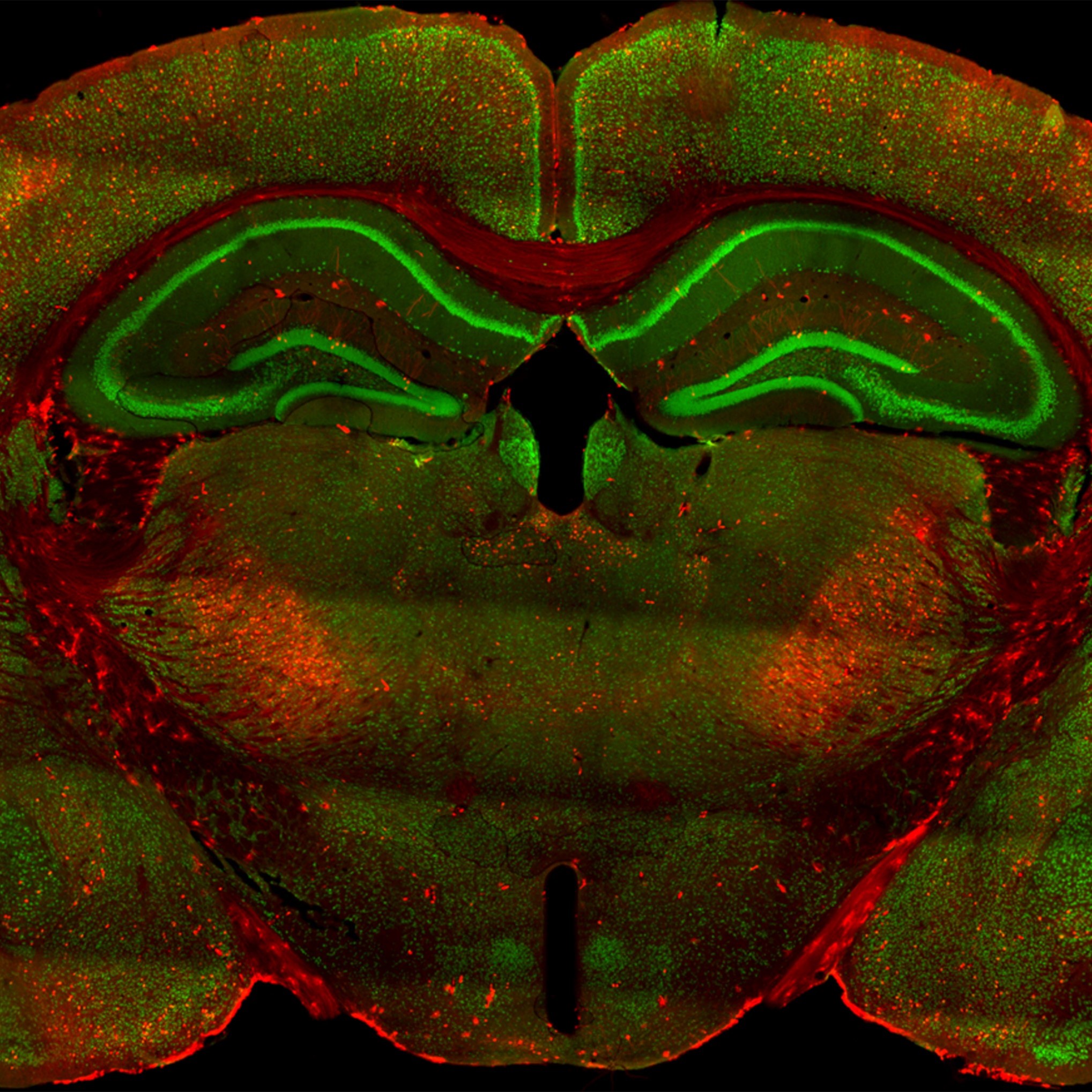
Confocal image of the hippocampus showing somatostatin inhibitory neurons (green).
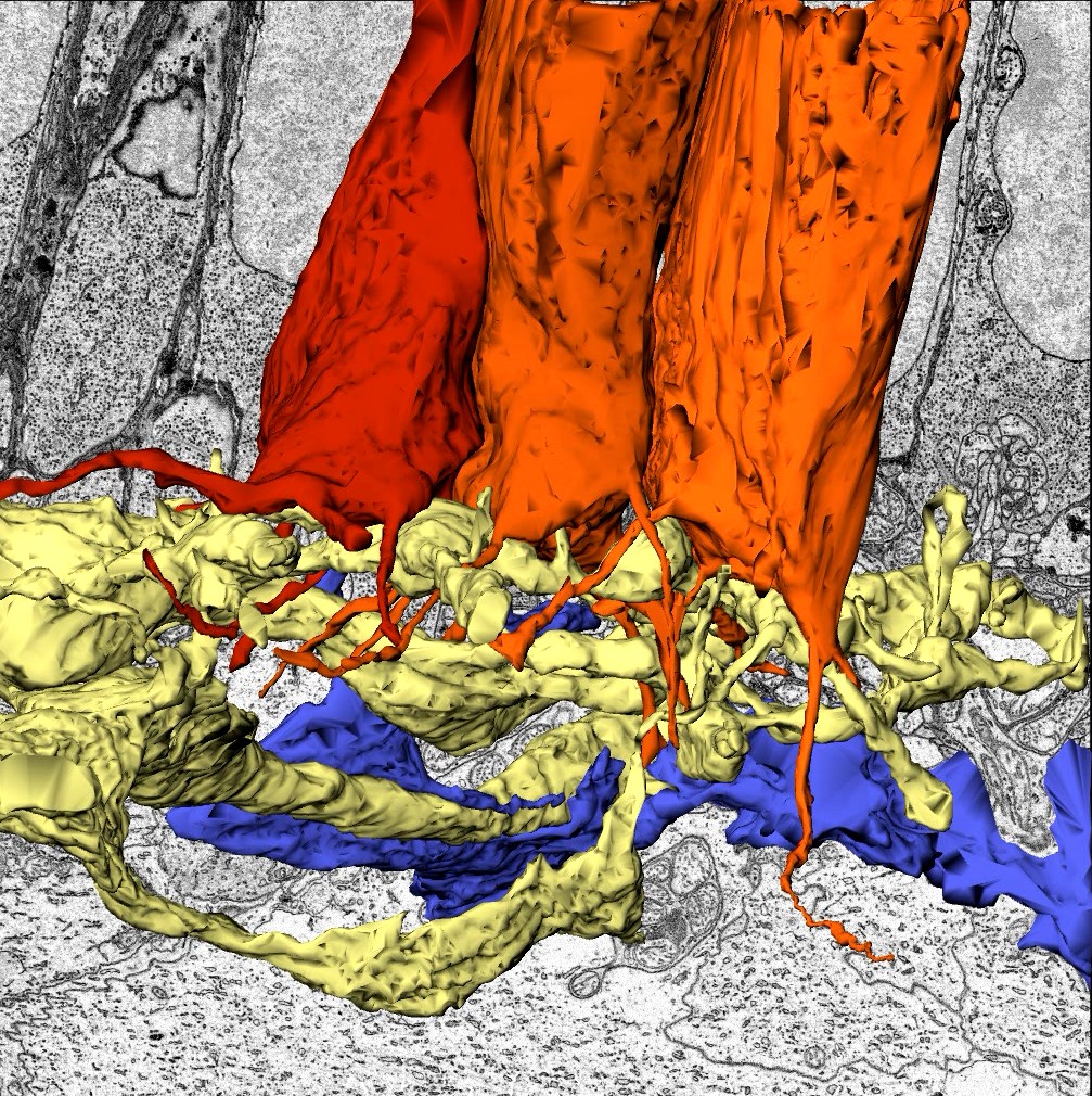
This image shows 3D reconstructions of Little skate photoreceptor terminals (orange) and postsynaptic partners (yellow and blue) obtained from serial block-face electron microscopy data.
Confocal image of a brain section containing the somatosensory (barrel) cortex from a transgenic mouse.
Green cells in this image are neurons distributed in the superficial region of the cerebral neocortex at postnatal day 9 in a heterozygous mutant mouse of Dab1.
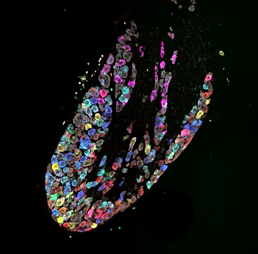
A composite of 5 images captured following each of 5 rounds of labeling and reprobing for a total of 12 mRNA transcripts and neuronal marker.
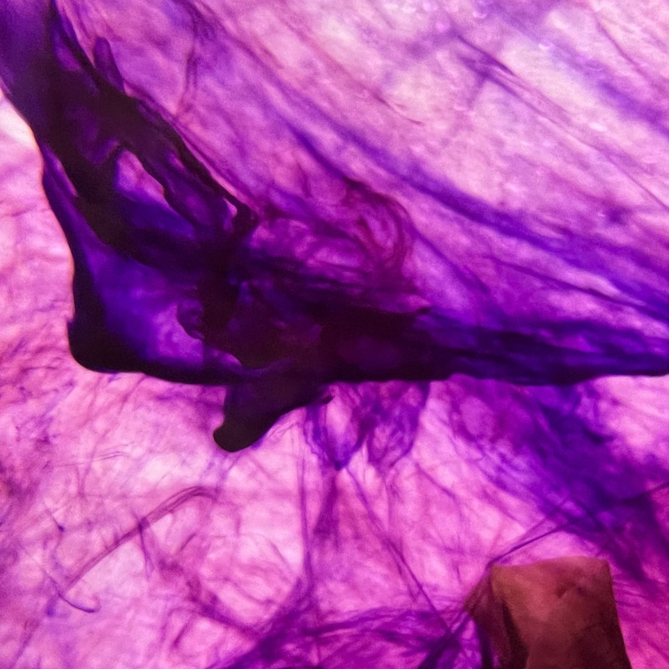
This image shows the inking response from a marine sea slug, Aplysia california, during sensitization training.
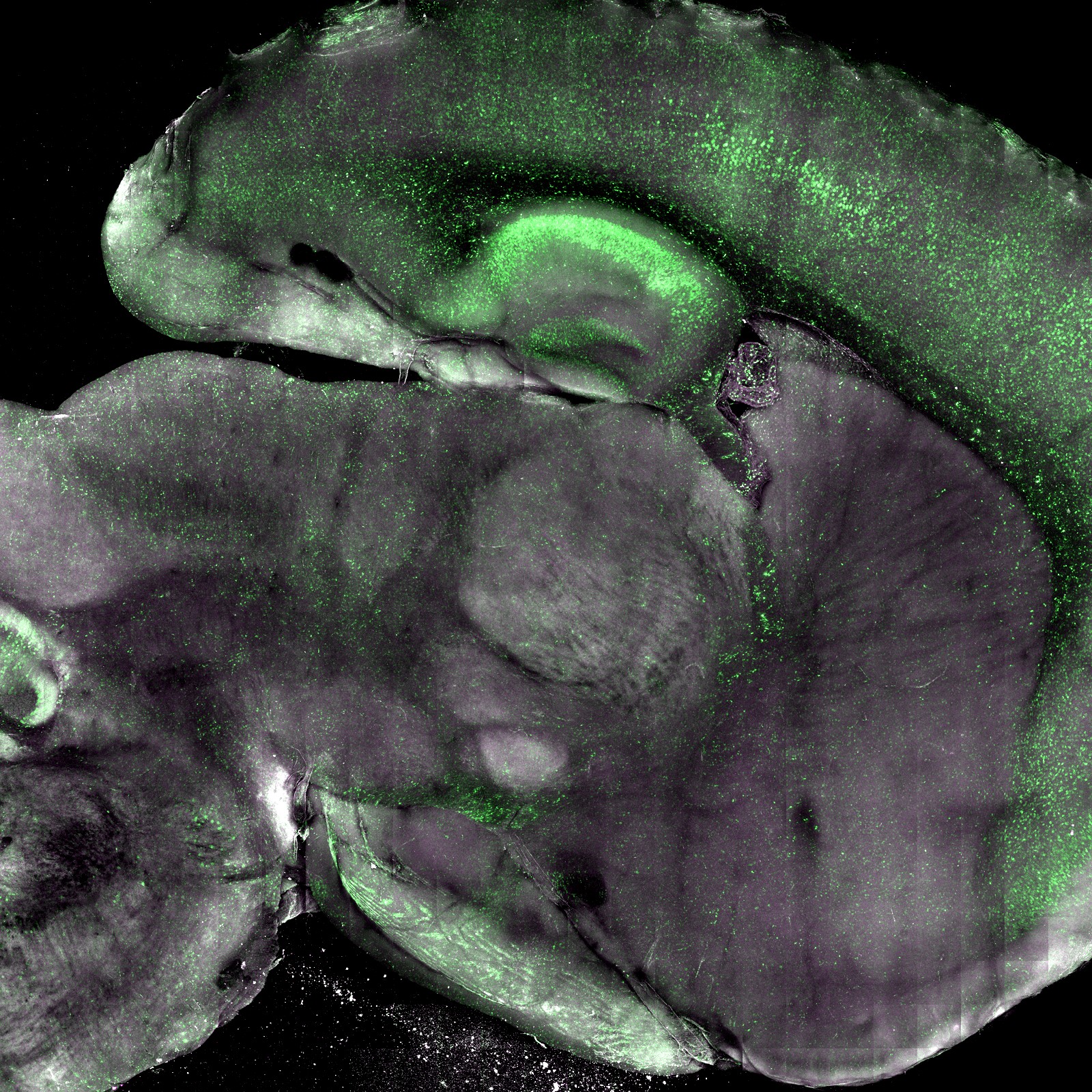
The image shows a clarified, 1-mm-thick sagittal section of whole brain from a transgenic mouse expressing a fluorescent reporter for caspase-3/7 activity.
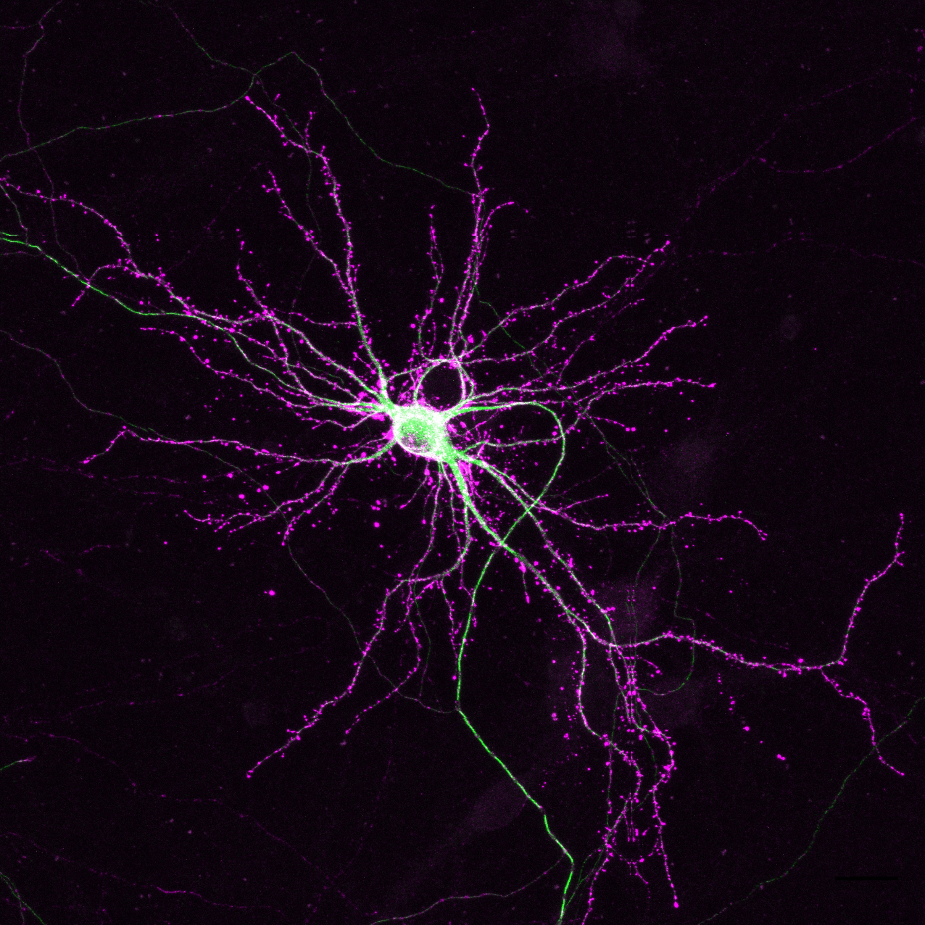
This image shows a hippocampal neuron with endogenous labelling of the cytoskeleton proteins β-actin and β3 tubulin using CRISPR/Cas9.
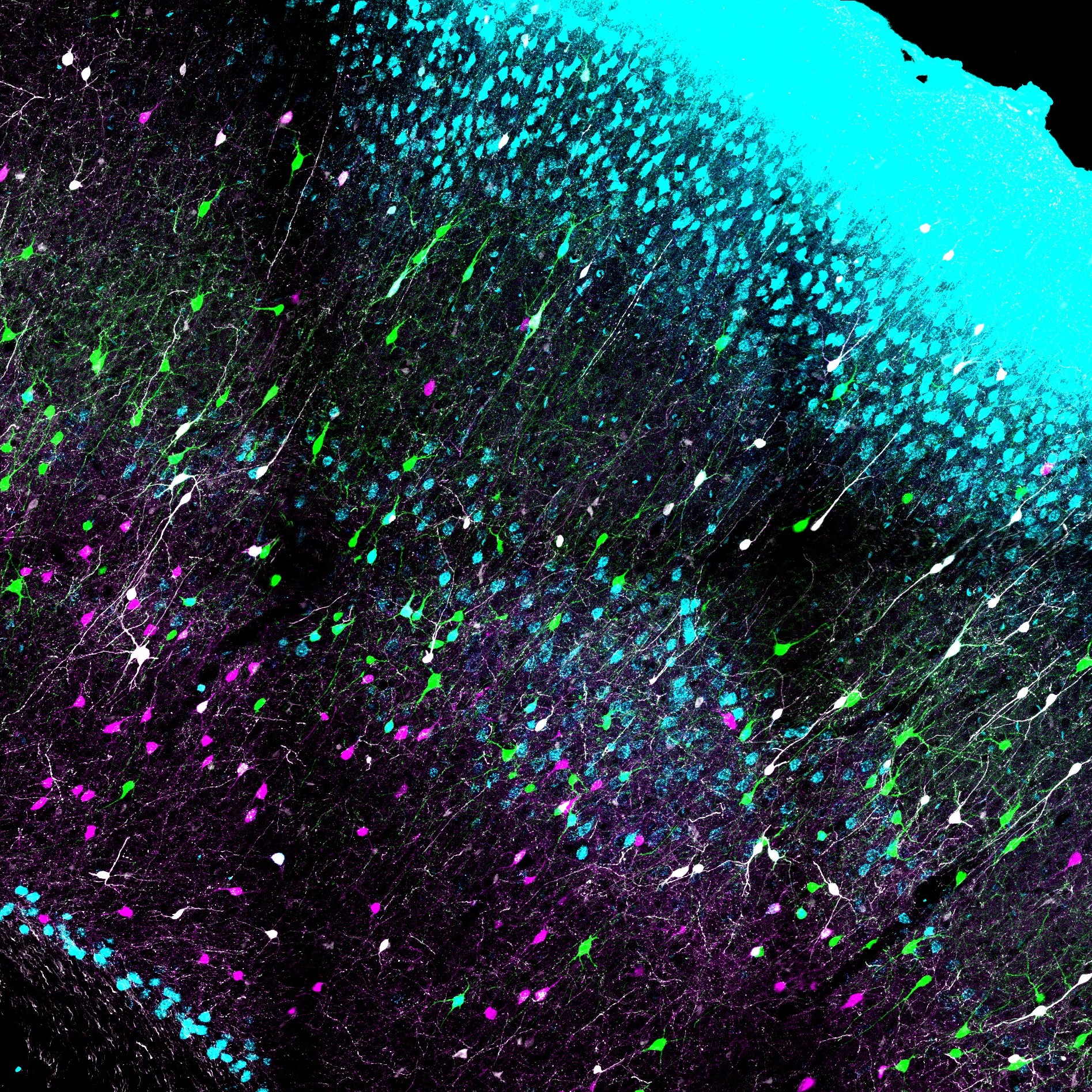
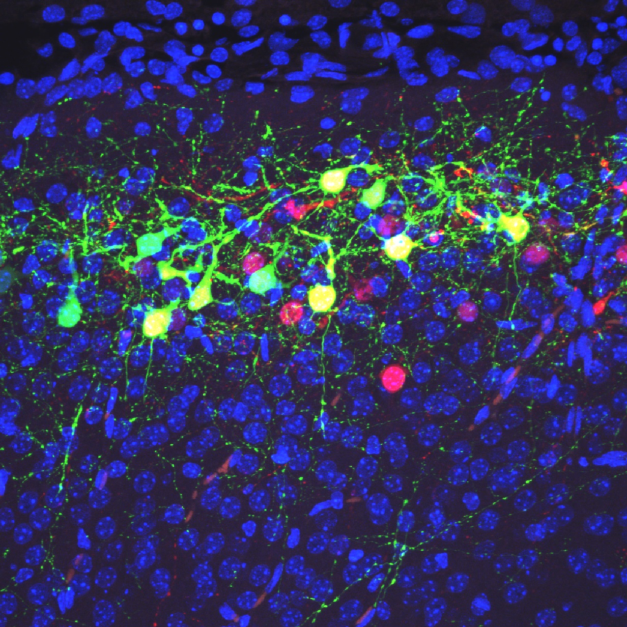
This image shows coronal slices at the level of the hippocampus in TRAP2 mice labeling neuronal ensembles during a rewarded T-maze spatial memory task.
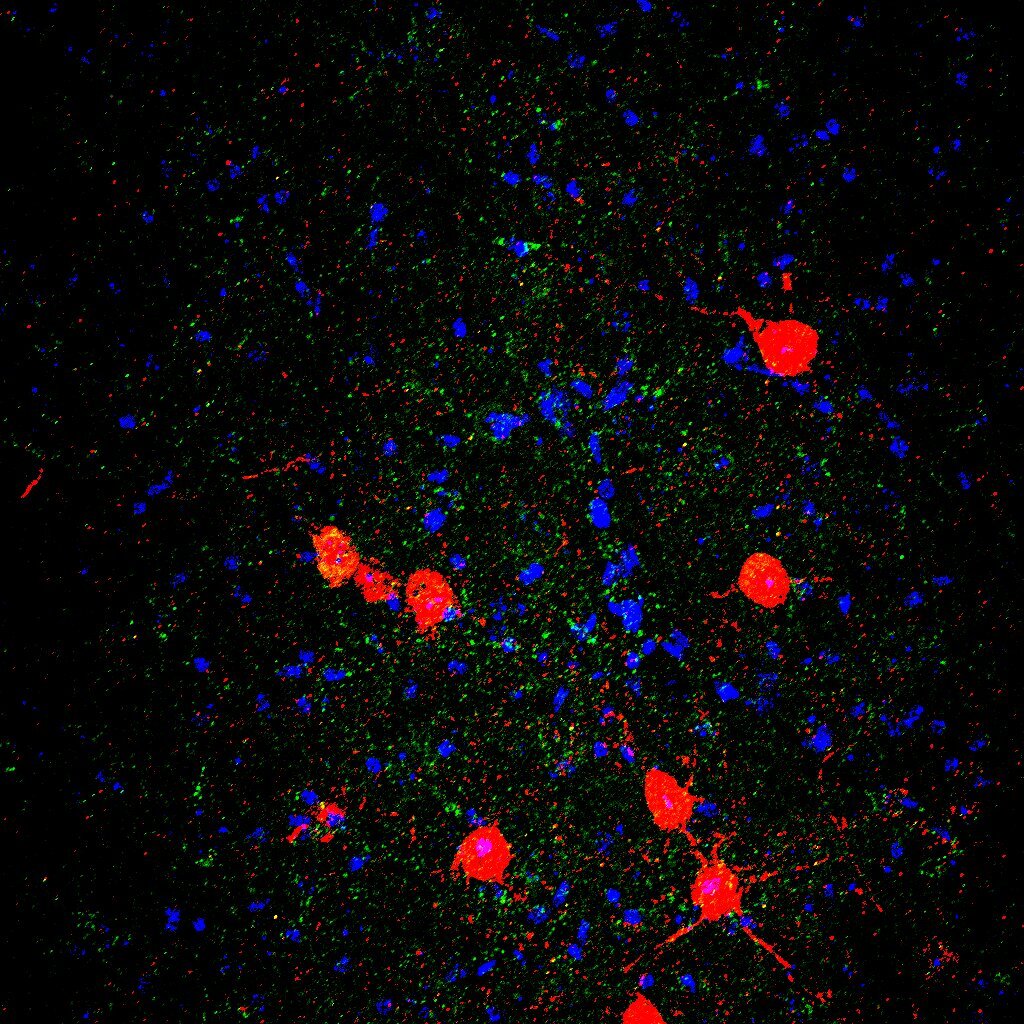
These images show the GABAA receptor δ subunit expressed on parvalbumin-positive interneurons in the basolateral amygdala.
FOLLOW US
 RSS Feed
TAGS
CATEGORIES


