Snapshots in Neuroscience
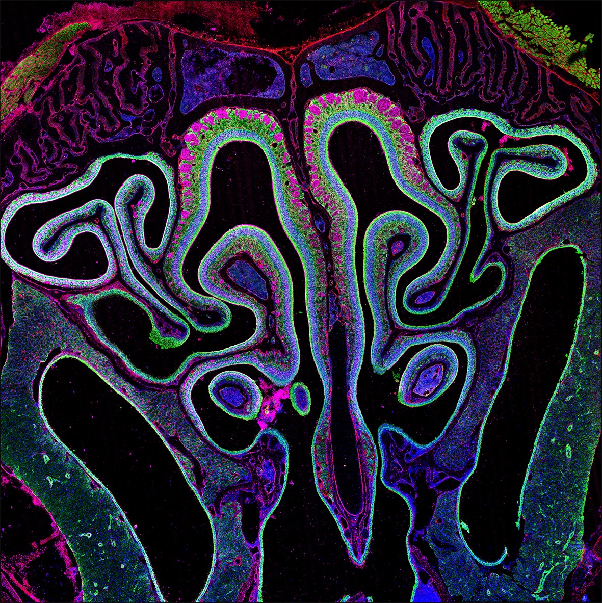
Shown are olfactory sensory neurons, sustentacular cells, and scattered sustentacular cells and macrophages infected with SARS-CoV-2.
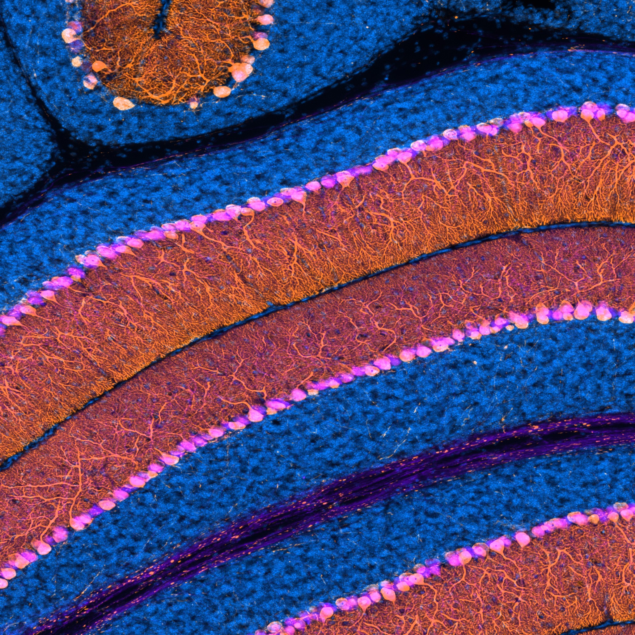
Visible in this sagittal section of a mouse cerebellum are granule cells and Purkinje cells.
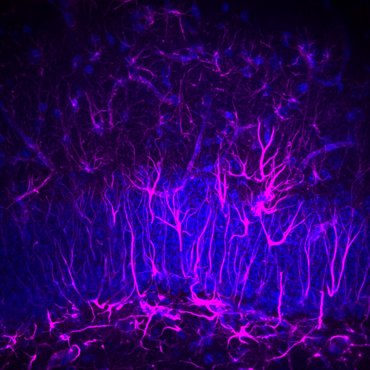
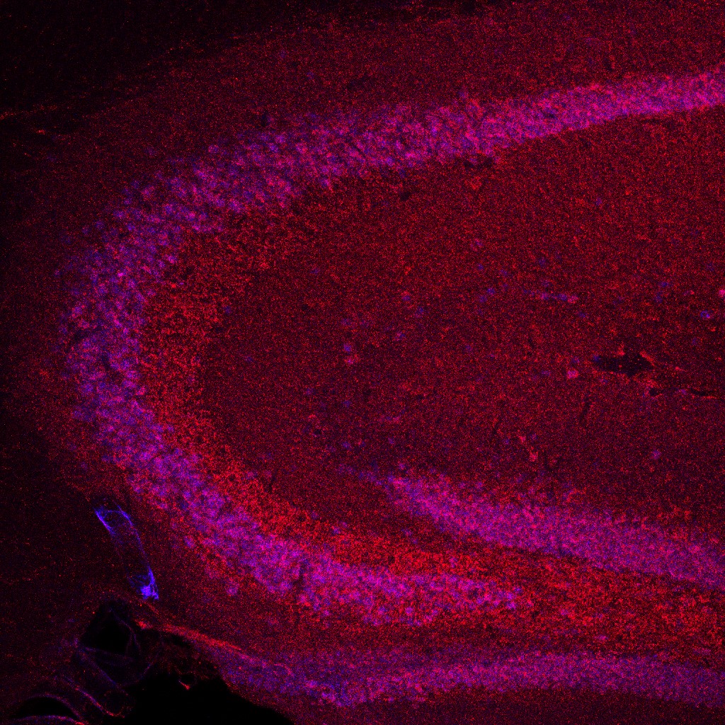
Magenta GFAP staining shows that mouse hippocampal astrocytes have a typical swept-spider morphology, while radial glial cells extend radially through the granule cell layer.
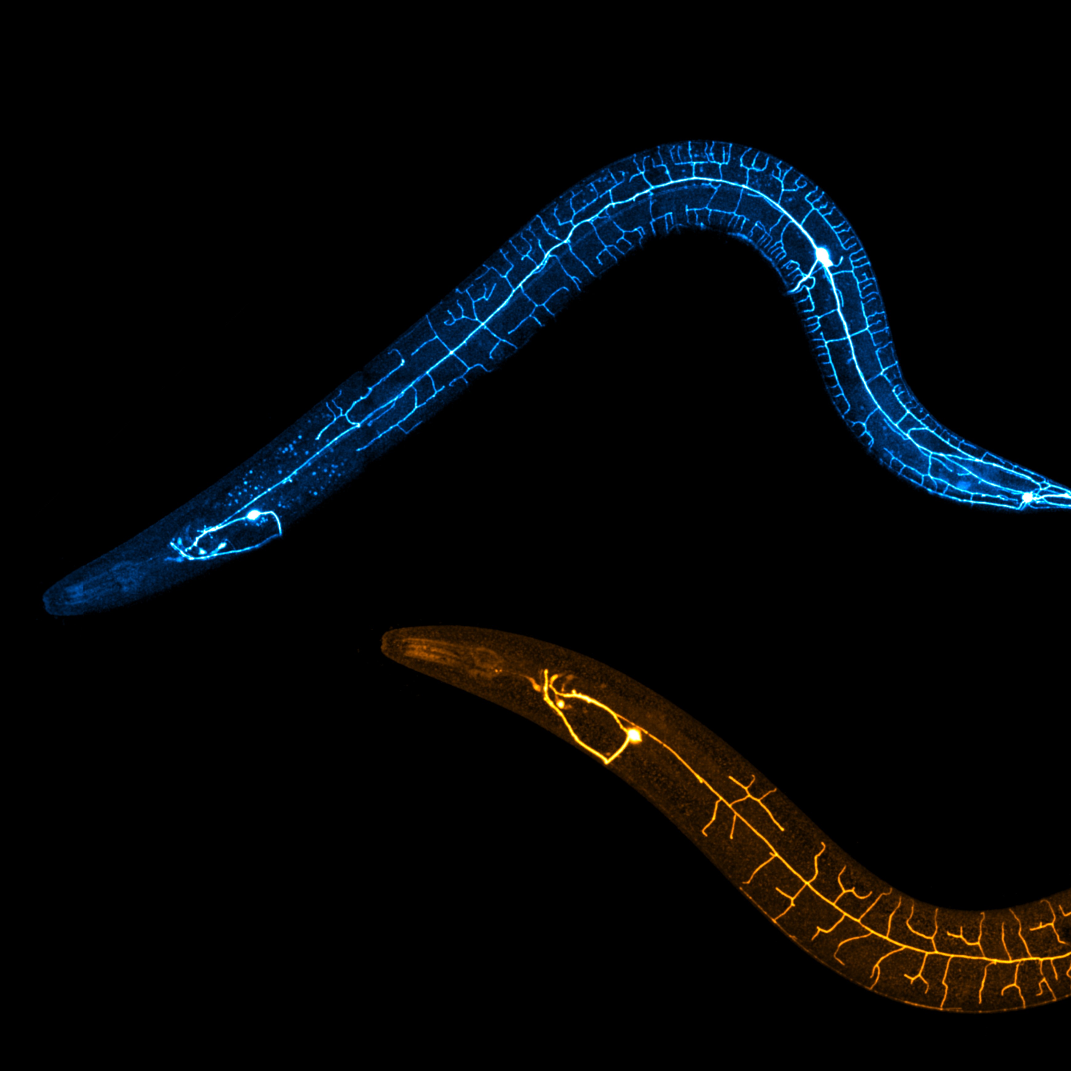
Shown are regenerating dendrites and uninjured dendrites of PVD neurons in C. elegans.
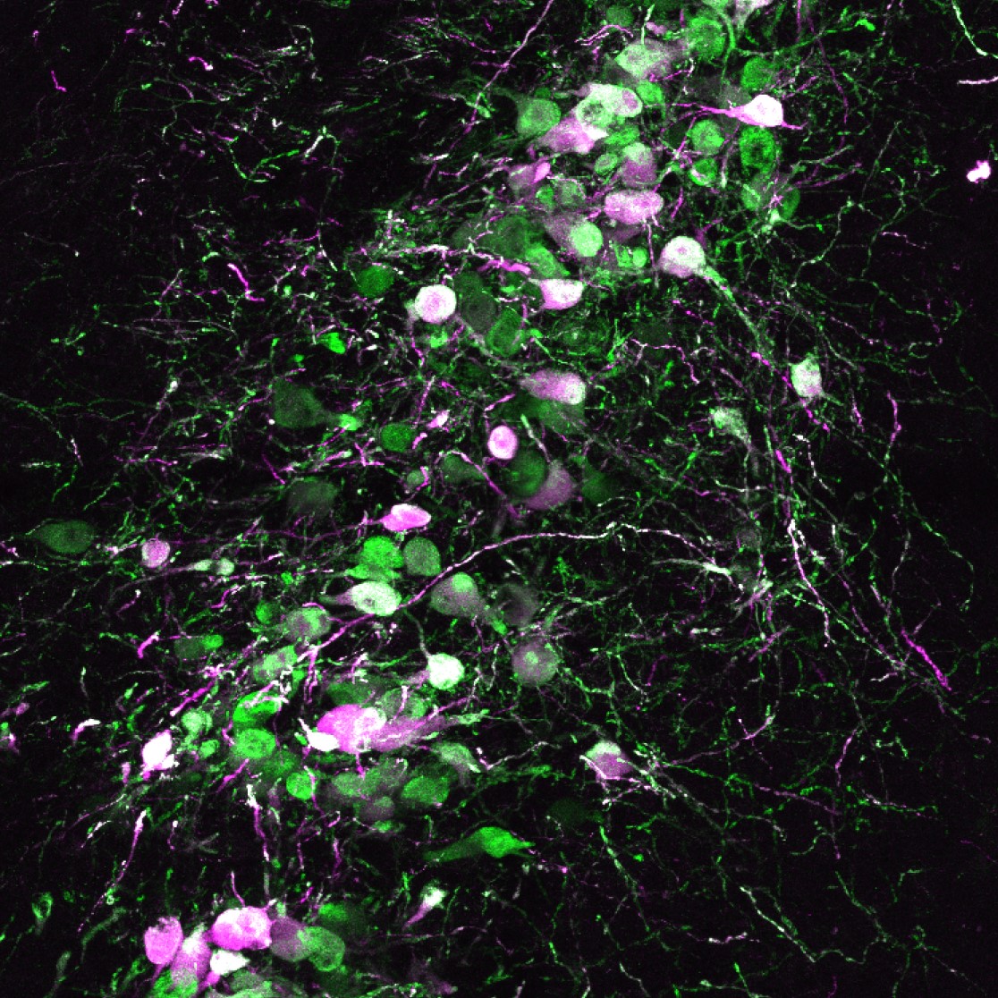
To assess neural activity during mouse tasting behavior, a calcium indicator (green) and a red fluorescent protein (magenta) were expressed in the locus coeruleus.
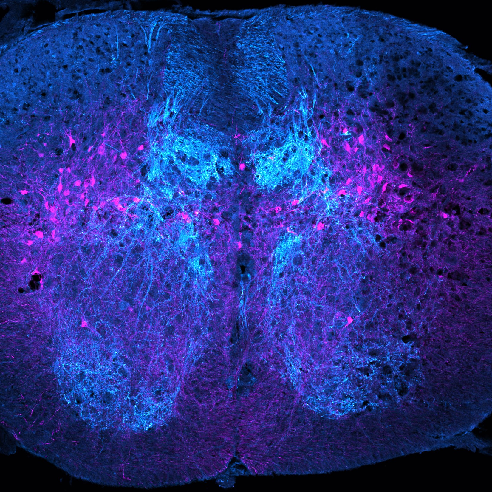
Pictured are proprioceptive sensory afferents and Atoh1-lineage neurons in the lower thoracic mouse spinal cord.
Reconstructed 3-dimensional whole brain distribution of orexin receptor expression visualized using the branched hybridization chain reaction method.
Two images of tree shrew retina captured with in vivo optical coherence tomography and ex vivo confocal imaging reveal densely packed, vertically elongated, and stratified axon bundles that are more like axon bundles in humans than in mice.
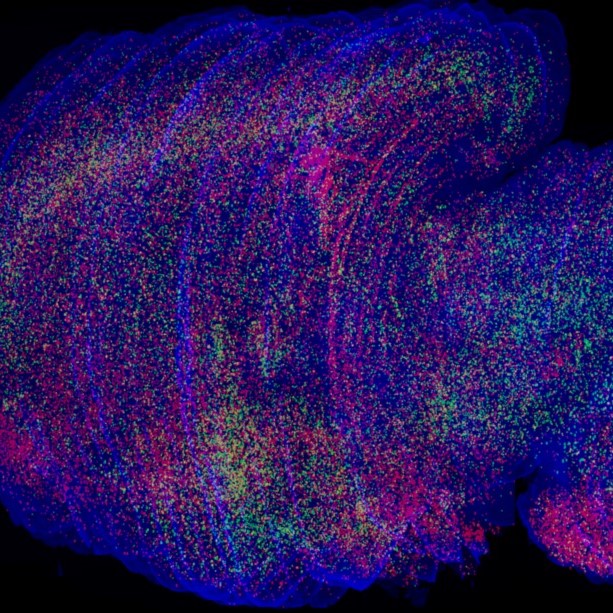
A maximum intensity projection image of a sagittal mouse brain slice captured using confocal microscopy.
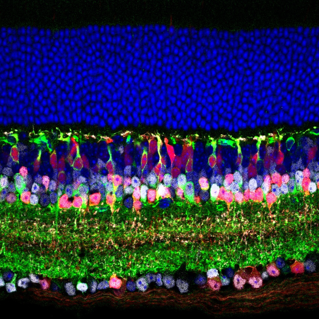
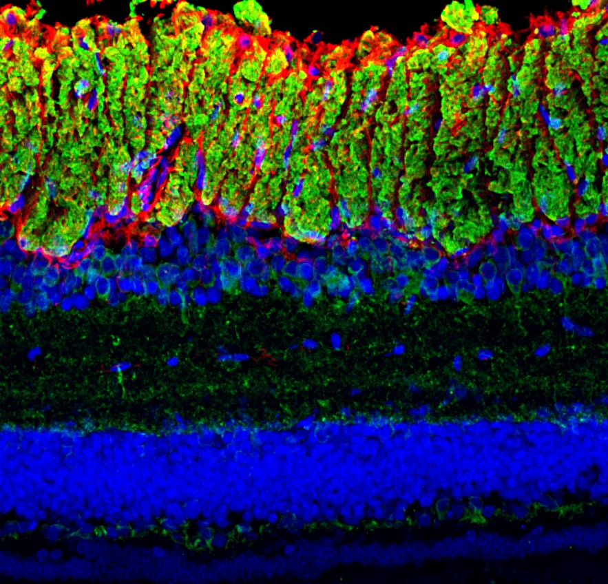
This image shows the cellular layers of an adult mouse retina, stained for markers of amacrine cells and type 3b bipolar cells.
FOLLOW US
 RSS Feed
TAGS
CATEGORIES


