Development
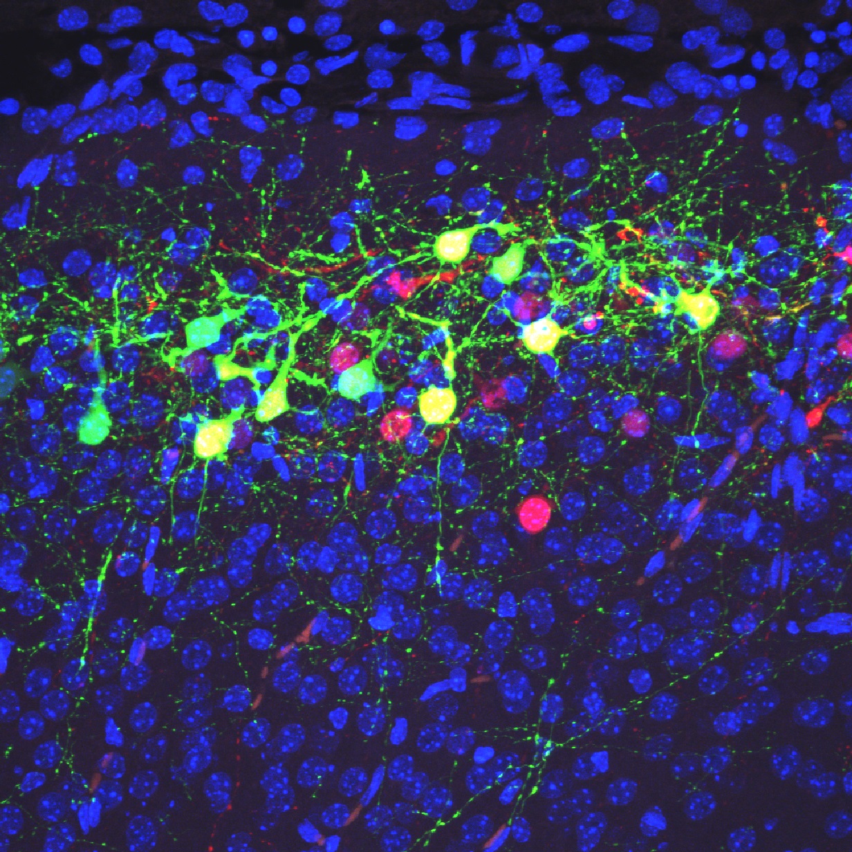
Green cells in this image are neurons distributed in the superficial region of the cerebral neocortex at postnatal day 9 in a heterozygous mutant mouse of Dab1.
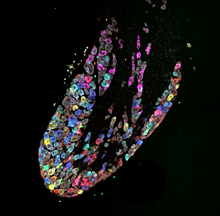
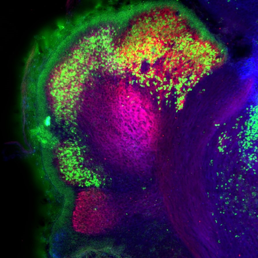
A composite of 5 images captured following each of 5 rounds of labeling and reprobing for a total of 12 mRNA transcripts and neuronal marker.
This image shows the developing central nervous system in a Drosophila embryo.
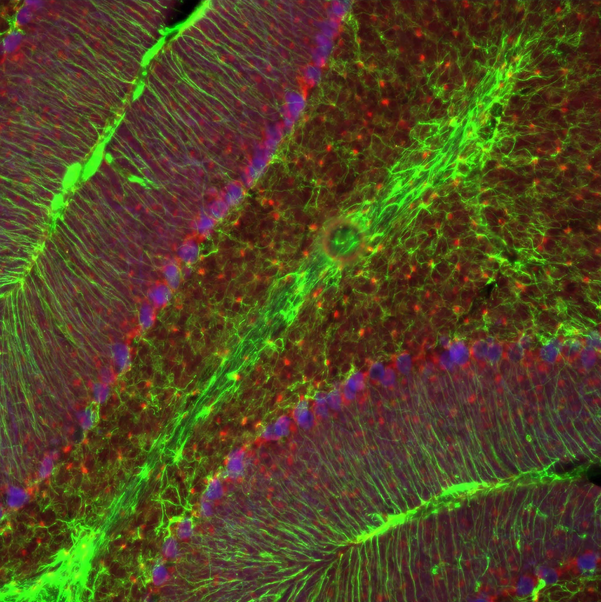
Part of the juvenile cerebellar cortex of a mouse at postnatal day 21.
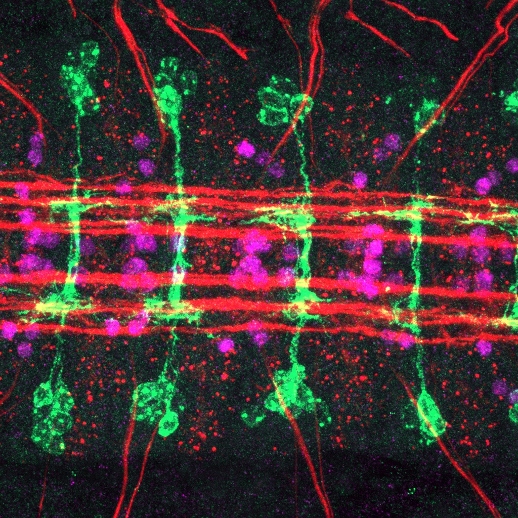
Purkinje cell clusters in the mouse cerebellum at embryonic day 17.5.
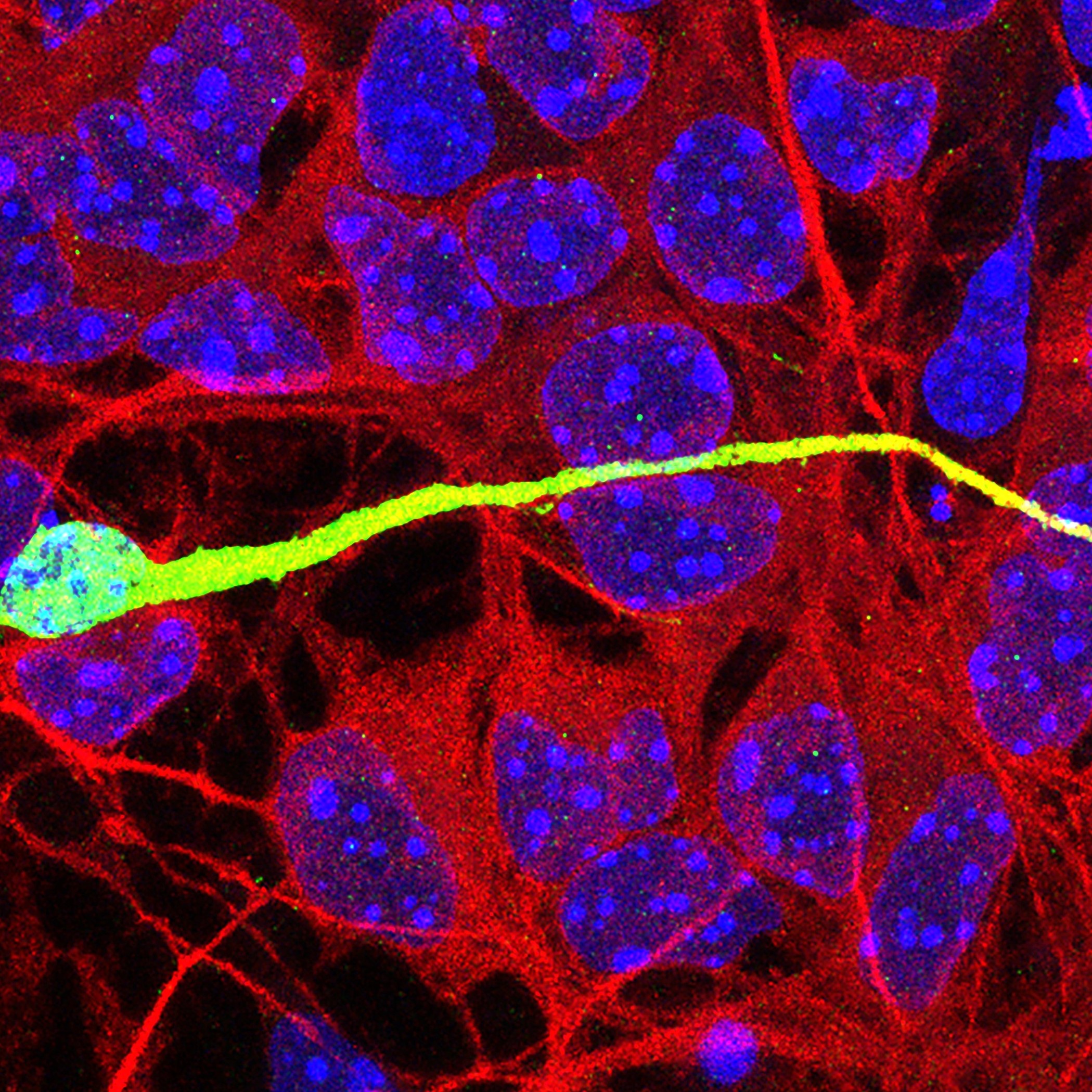
This image shows an interneuron migrating on a monolayer of cortical feeder cells in an in vitro co-culture assay.
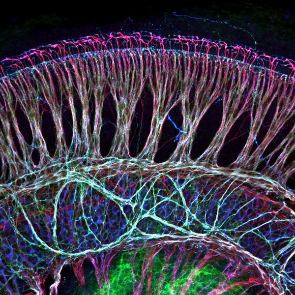
This image shows an immunostained whole-mount preparation of a mouse cochlea.
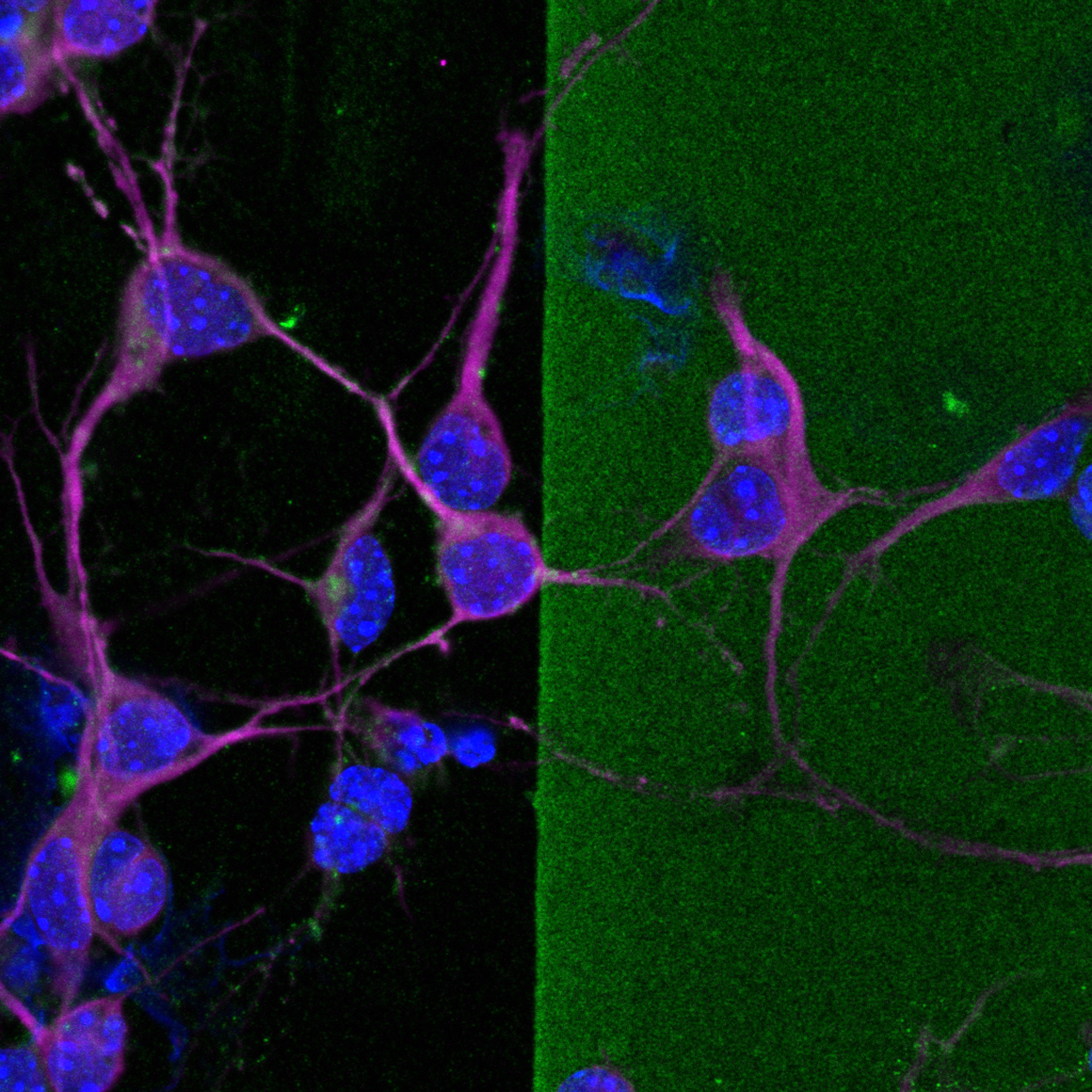
This image shows MAP2+ neurites from cultured immature cortical neurons extending into a stripe of chondroitin sulfate proteoglycans (CSPGs) treated with chondroitinase ABC, an enzyme that abolishes CSPGs
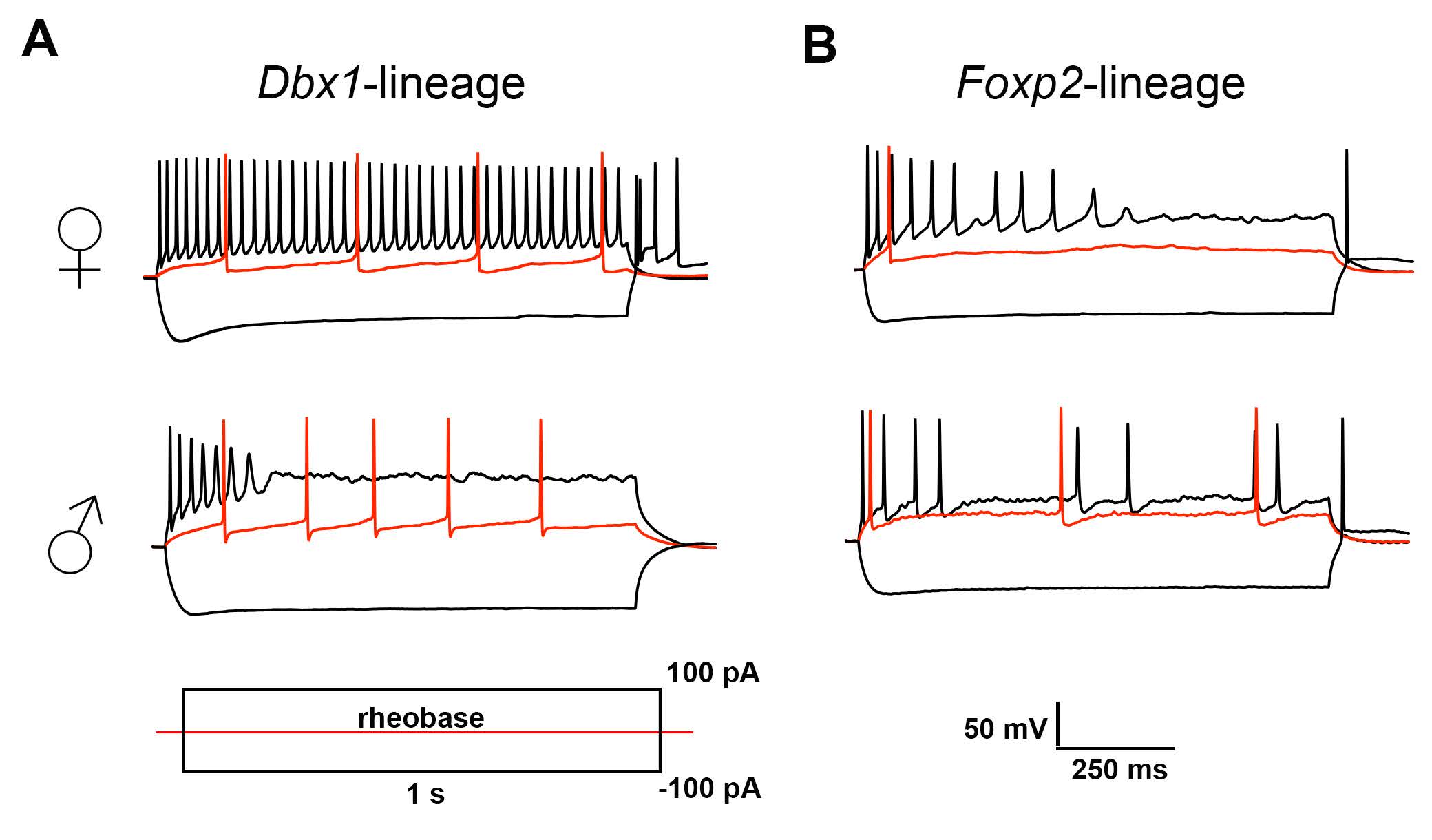
Authors show neuronal subpopulation and sex differences in the biophysical signatures of developmentally defined medial amygdala output neurons.
Authors examined the reliability of using Cre-induced recombination of one gene to predict recombination in another gene at the single-cell level in adult hippocampal neural stem and progenitor cells.
FOLLOW US
 RSS Feed
TAGS
CATEGORIES


