Snapshots in Neuroscience
Excitatory axon terminals expressing vesicular glutamate transporters in the thalamus of a tree shrew.
Real-time calcium dynamics of astrocytes and oxytocin neurons in the mouse paraventricular nucleus of the hypothalamus.

Exploring the neuronal expression of seizure-associated proteins in the fruit fly brain.
New mouse model for inducing targeted gene expression within neurons of the spinal cord dorsal horn.
Immunostaining of cell bodies and nerve fibers of neurons in an adult mouse small intestinal myenteric ganglion.
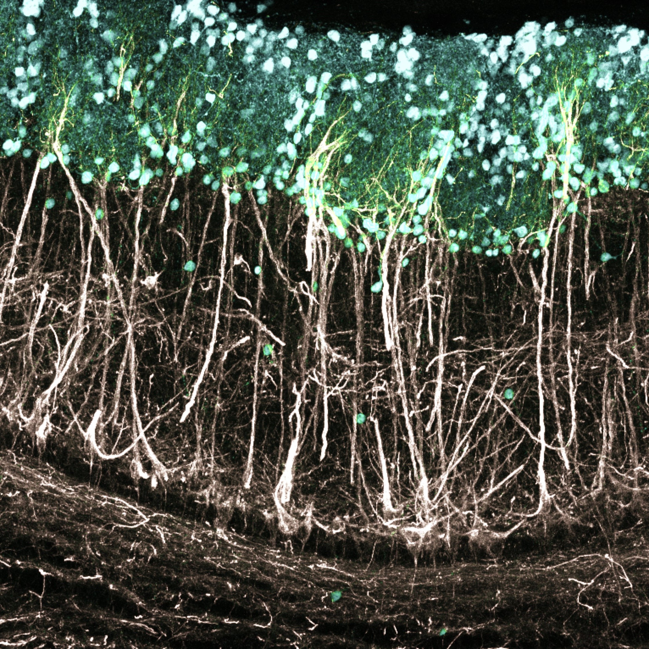
Principal neuron dendrites in the adult mouse olfactory bulb extending up towards the glomerular layer where they receive sensory input.
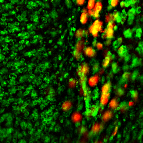
Coronal section of a mouse nucleus accumbens with GABAergic neurons ablated.
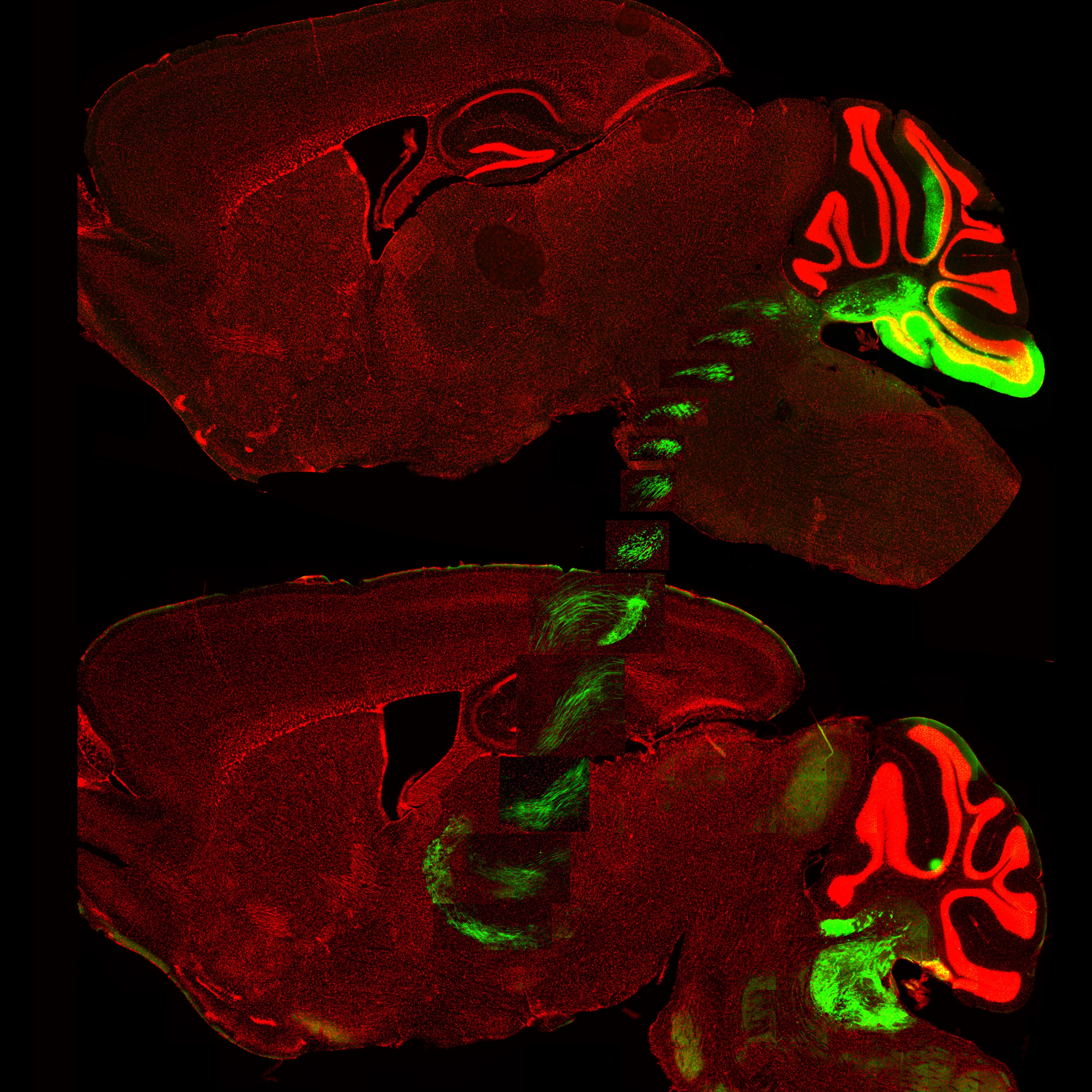
Sagittal mouse brain sections showing cerebellar nuclei sending afferents to the ventral posterolateral nucleus of the thalamus.
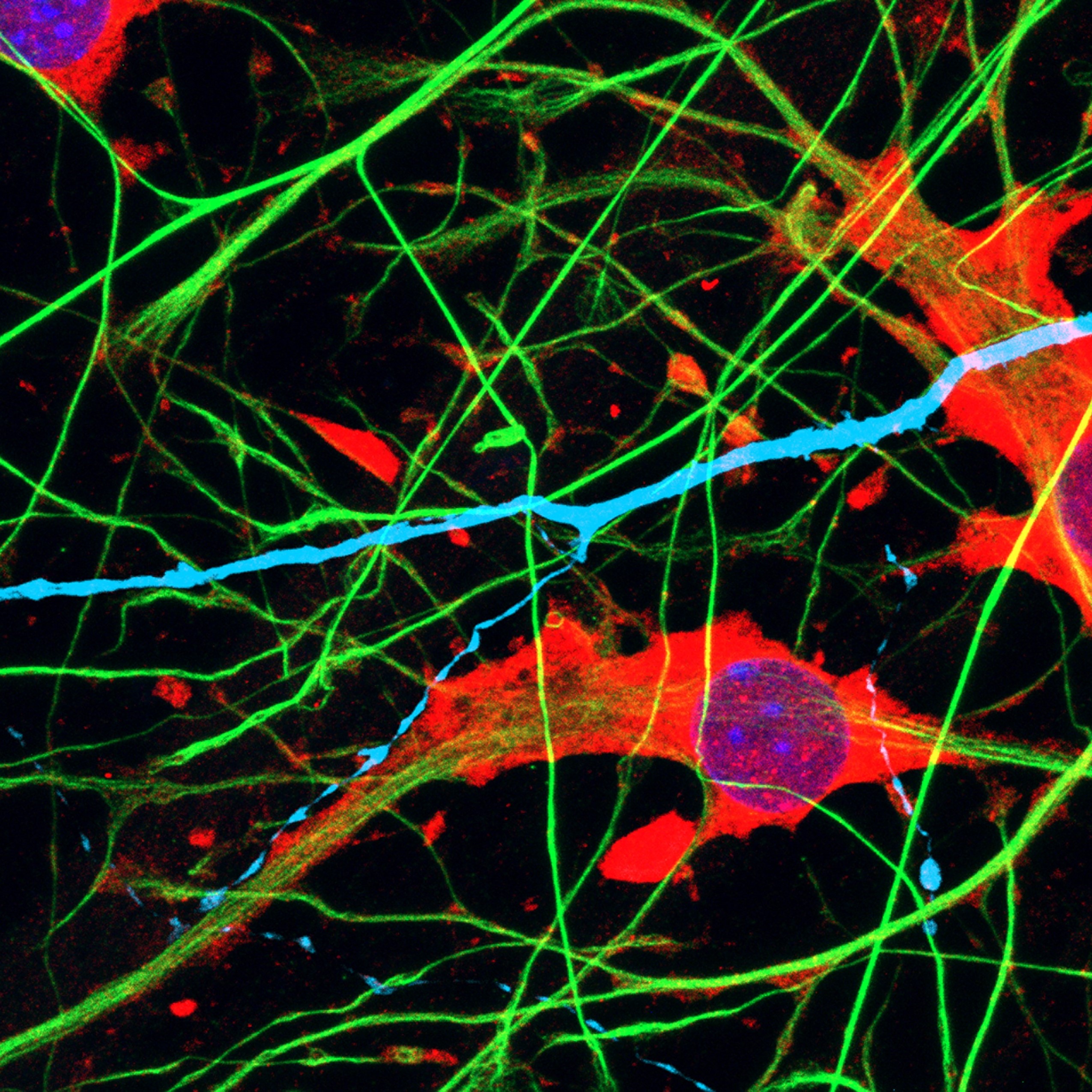
Cocultured human induced pluripotent stem cell-derived astrocytes and neurons.
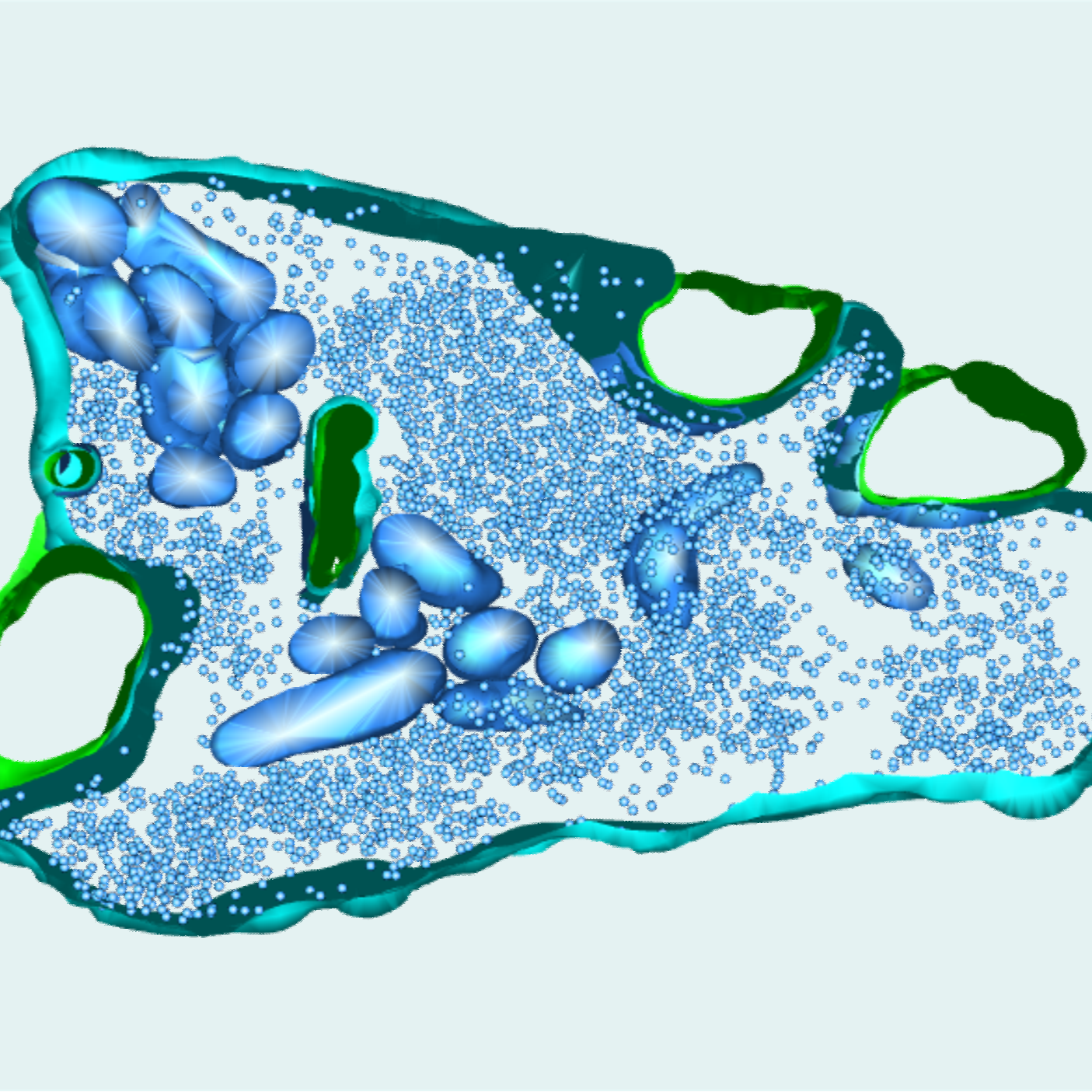
These partial 3D reconstructions of synapses from wild-type and synapsin knock-out mice show the presynaptic membrane, the postsynaptic membrane, and synaptic vesicles.
FOLLOW US
 RSS Feed
TAGS
CATEGORIES


